POLCAM: instant molecular orientation microscopy for the life sciences
IF 36.1
1区 生物学
Q1 BIOCHEMICAL RESEARCH METHODS
引用次数: 0
Abstract
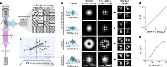
Current methods for single-molecule orientation localization microscopy (SMOLM) require optical setups and algorithms that can be prohibitively slow and complex, limiting widespread adoption for biological applications. We present POLCAM, a simplified SMOLM method based on polarized detection using a polarization camera, which can be easily implemented on any wide-field fluorescence microscope. To make polarization cameras compatible with single-molecule detection, we developed theory to minimize field-of-view errors, used simulations to optimize experimental design and developed a fast algorithm based on Stokes parameter estimation that can operate over 1,000-fold faster than the state of the art, enabling near-instant determination of molecular anisotropy. To aid in the adoption of POLCAM, we developed open-source image analysis software and a website detailing hardware installation and software use. To illustrate the potential of POLCAM in the life sciences, we applied our method to study α-synuclein fibrils, the actin cytoskeleton of mammalian cells, fibroblast-like cells and the plasma membrane of live human T cells. Combining localization and polarization microscopy can yield detailed insights into subcellular structures. POLCAM uses a polarization camera and wide-field microscopy for rapid measurement of super-resolution orientation imaging in live cells.

POLCAM:用于生命科学的即时分子定向显微镜。
目前的单分子定向定位显微镜(SMOLM)方法需要的光学设置和算法可能过于缓慢和复杂,从而限制了生物应用的广泛采用。我们提出的 POLCAM 是一种简化的 SMOLM 方法,它基于使用偏振相机的偏振检测,可在任何宽视场荧光显微镜上轻松实现。为了使偏振相机与单分子检测兼容,我们开发了将视场误差最小化的理论,使用模拟来优化实验设计,并开发了基于斯托克斯参数估计的快速算法,该算法的运行速度比现有技术快 1000 多倍,可近乎即时地确定分子各向异性。为了帮助 POLCAM 的应用,我们开发了开源图像分析软件和一个详细介绍硬件安装和软件使用的网站。为了说明 POLCAM 在生命科学领域的潜力,我们应用我们的方法研究了 α-突触核蛋白纤维、哺乳动物细胞的肌动蛋白细胞骨架、类成纤维细胞和活人 T 细胞的质膜。
本文章由计算机程序翻译,如有差异,请以英文原文为准。
求助全文
约1分钟内获得全文
求助全文
来源期刊

Nature Methods
生物-生化研究方法
CiteScore
58.70
自引率
1.70%
发文量
326
审稿时长
1 months
期刊介绍:
Nature Methods is a monthly journal that focuses on publishing innovative methods and substantial enhancements to fundamental life sciences research techniques. Geared towards a diverse, interdisciplinary readership of researchers in academia and industry engaged in laboratory work, the journal offers new tools for research and emphasizes the immediate practical significance of the featured work. It publishes primary research papers and reviews recent technical and methodological advancements, with a particular interest in primary methods papers relevant to the biological and biomedical sciences. This includes methods rooted in chemistry with practical applications for studying biological problems.
 求助内容:
求助内容: 应助结果提醒方式:


