Molecular basis of plastoquinone reduction in plant cytochrome b6f

IF 15.8 1区 生物学 Q1 PLANT SCIENCES
Sebastian Pintscher, Rafał Pietras, Bohun Mielecki, Mateusz Szwalec, Anna Wójcik-Augustyn, Paulina Indyka, Michał Rawski, Łukasz Koziej, Marcin Jaciuk, Grzegorz Ważny, Sebastian Glatt, Artur Osyczka
{"title":"Molecular basis of plastoquinone reduction in plant cytochrome b6f","authors":"Sebastian Pintscher, Rafał Pietras, Bohun Mielecki, Mateusz Szwalec, Anna Wójcik-Augustyn, Paulina Indyka, Michał Rawski, Łukasz Koziej, Marcin Jaciuk, Grzegorz Ważny, Sebastian Glatt, Artur Osyczka","doi":"10.1038/s41477-024-01804-x","DOIUrl":null,"url":null,"abstract":"<p>A multi-subunit enzyme, cytochrome <i>b</i><sub>6</sub><i>f</i> (cyt<i>b</i><sub>6</sub><i>f</i>), provides the crucial link between photosystems I and II in the photosynthetic membranes of higher plants, transferring electrons between plastoquinone (PQ) and plastocyanin. The atomic structure of cyt<i>b</i><sub>6</sub><i>f</i> is known, but its detailed catalytic mechanism remains elusive. Here we present cryogenic electron microscopy structures of spinach cyt<i>b</i><sub>6</sub><i>f</i> at 1.9 Å and 2.2 Å resolution, revealing an unexpected orientation of the substrate PQ in the haem ligand niche that forms the PQ reduction site (Q<sub>n</sub>). PQ, unlike Q<sub>n</sub> inhibitors, is not in direct contact with the haem. Instead, a water molecule is coordinated by one of the carbonyl groups of PQ and can act as the immediate proton donor for PQ. In addition, we identify water channels that connect Q<sub>n</sub> with the aqueous exterior of the enzyme, suggesting that the binding of PQ in Q<sub>n</sub> displaces water through these channels. The structures confirm large movements of the head domain of the iron–sulfur protein (ISP-HD) towards and away from the plastoquinol oxidation site (Q<sub>p</sub>) and define the unique position of ISP-HD when a Q<sub>p</sub> inhibitor (2,5-dibromo-3-methyl-6-isopropylbenzoquinone) is bound. This work identifies key conformational states of cyt<i>b</i><sub>6</sub><i>f</i>, highlights fundamental differences between substrates and inhibitors and proposes a quinone–water exchange mechanism.</p>","PeriodicalId":18904,"journal":{"name":"Nature Plants","volume":null,"pages":null},"PeriodicalIF":15.8000,"publicationDate":"2024-10-03","publicationTypes":"Journal Article","fieldsOfStudy":null,"isOpenAccess":false,"openAccessPdf":"","citationCount":"0","resultStr":null,"platform":"Semanticscholar","paperid":null,"PeriodicalName":"Nature Plants","FirstCategoryId":"99","ListUrlMain":"https://doi.org/10.1038/s41477-024-01804-x","RegionNum":1,"RegionCategory":"生物学","ArticlePicture":[],"TitleCN":null,"AbstractTextCN":null,"PMCID":null,"EPubDate":"","PubModel":"","JCR":"Q1","JCRName":"PLANT SCIENCES","Score":null,"Total":0}
引用次数: 0

Abstract

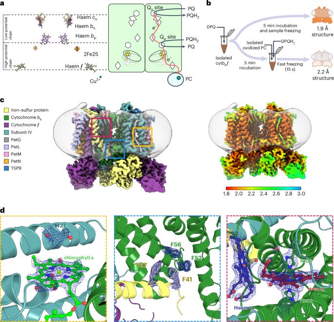
A multi-subunit enzyme, cytochrome b6f (cytb6f), provides the crucial link between photosystems I and II in the photosynthetic membranes of higher plants, transferring electrons between plastoquinone (PQ) and plastocyanin. The atomic structure of cytb6f is known, but its detailed catalytic mechanism remains elusive. Here we present cryogenic electron microscopy structures of spinach cytb6f at 1.9 Å and 2.2 Å resolution, revealing an unexpected orientation of the substrate PQ in the haem ligand niche that forms the PQ reduction site (Qn). PQ, unlike Qn inhibitors, is not in direct contact with the haem. Instead, a water molecule is coordinated by one of the carbonyl groups of PQ and can act as the immediate proton donor for PQ. In addition, we identify water channels that connect Qn with the aqueous exterior of the enzyme, suggesting that the binding of PQ in Qn displaces water through these channels. The structures confirm large movements of the head domain of the iron–sulfur protein (ISP-HD) towards and away from the plastoquinol oxidation site (Qp) and define the unique position of ISP-HD when a Qp inhibitor (2,5-dibromo-3-methyl-6-isopropylbenzoquinone) is bound. This work identifies key conformational states of cytb6f, highlights fundamental differences between substrates and inhibitors and proposes a quinone–water exchange mechanism.

Abstract Image

植物细胞色素 b6f 中质子醌还原的分子基础
一种多亚基酶--细胞色素 b6f(cytb6f)--是高等植物光合膜中光合系统 I 和 II 之间的关键纽带,它在质醌(PQ)和质花青素之间传递电子。细胞b6f的原子结构已知,但其详细的催化机理仍不清楚。在这里,我们以 1.9 Å 和 2.2 Å 的分辨率展示了菠菜 cytb6f 的低温电子显微镜结构,揭示了底物 PQ 在形成 PQ 还原位点(Qn)的血红素配体位点中的意外取向。与 Qn 抑制剂不同,PQ 并不与血红素直接接触。相反,一个水分子与 PQ 的一个羰基配位,可作为 PQ 的直接质子供体。此外,我们还发现了连接 Qn 与酶的水外部的水通道,这表明 Qn 中 PQ 的结合会通过这些通道置换出水。这些结构证实了铁硫蛋白头部结构域(ISP-HD)向和远离质醌氧化位点(Qp)的大量运动,并确定了当 Qp 抑制剂(2,5-二溴-3-甲基-6-异丙基苯醌)结合时 ISP-HD 的独特位置。这项工作确定了细胞b6f的关键构象状态,突出了底物和抑制剂之间的根本差异,并提出了一种醌水交换机制。
本文章由计算机程序翻译,如有差异,请以英文原文为准。
求助全文
约1分钟内获得全文 求助全文
来源期刊
Nature Plants
Nature Plants PLANT SCIENCES-
CiteScore
25.30
自引率
2.20%
发文量
196
期刊介绍: Nature Plants is an online-only, monthly journal publishing the best research on plants — from their evolution, development, metabolism and environmental interactions to their societal significance.
×
引用
GB/T 7714-2015
复制
MLA
复制
APA
复制
导出至
BibTeX EndNote RefMan NoteFirst NoteExpress
×
提示
您的信息不完整,为了账户安全,请先补充。
现在去补充
×
提示
您因"违规操作"
具体请查看互助需知
我知道了
×
提示
确定
请完成安全验证×
copy
已复制链接
快去分享给好友吧!
我知道了
右上角分享
点击右上角分享
0
联系我们:info@booksci.cn Book学术提供免费学术资源搜索服务,方便国内外学者检索中英文文献。致力于提供最便捷和优质的服务体验。 Copyright © 2023 布克学术 All rights reserved.
京ICP备2023020795号-1
ghs 京公网安备 11010802042870号
Book学术文献互助
Book学术文献互助群
群 号:481959085
Book学术官方微信