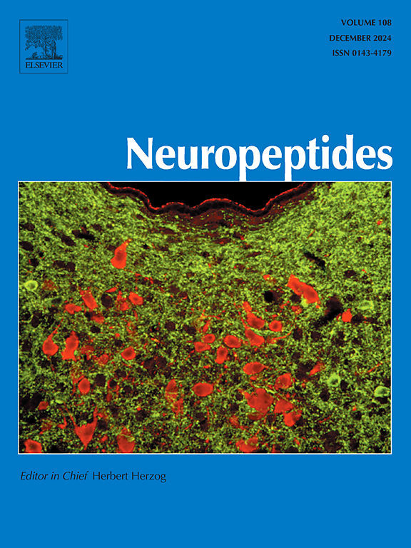
Phosphorylated NPY1R regulates phenotypic transition of vascular smooth muscle cells, inflammatory response and macrophage infiltration to promote intracranial aneurysm progression
IF 2.7
3区 医学
Q3 ENDOCRINOLOGY & METABOLISM
引用次数: 0
Abstract
Background
Rupture of intracranial aneurysm (IA) could give rise to spontaneous subarachnoid hemorrhage, leading to a high disability rate and even death. NPY1R expression was upregulated in aneurysm tissues of IA patients. However, the role and underlying mechanism of NPY1R remains unknown.
Methods
The IA model of mice was established using inducing systemic hypertension and injecting elastase. The expression of genes and proteins was detected by RT-qPCR and western blot. The number of T cells, macrophages, and neutrophils in IA mice was detected using flow cytometry and IF assay. The levels of inflammatory factors were measured using ELISA. Patho-morphology and inflammatory cells in aneurysm tissues were evaluated by HE staining. The interaction between TK and NPY1R was validated using Co-IP.
Results
NPY1R expression was greatly elevated in aneurysm tissues in IA patients and mice, which were positively related to macrophage infiltration. Besides, exogenous overexpression of NPY1R resulted in the promotion of contractile phenotype to the synthetic phenotype of vascular smooth muscle cells (VSMCs), inflammatory response and M1 macrophage polarization. In terms of the underlying mechanism, NPY1R protein could be modified by TK-mediated phosphorylation and TKI could decrease IA formation and suppresse contractile phenotype to synthetic phenotype of VSMCs, inflammatory response and M1 macrophage polarization in IA mice. Furthermore, ablating mouse macrophages abolished NPY1R overexpression-mediated promotion of IA formation and rupture in mice.
Conclusion
Phosphorylated NPY1R contributed to IA progression through promoting contractile phenotype to synthetic phenotype of VSMCs, inflammatory response and M1 macrophage polarization in IA.
磷酸化的 NPY1R 可调控血管平滑肌细胞的表型转换、炎症反应和巨噬细胞浸润,从而促进颅内动脉瘤的进展
背景颅内动脉瘤(IA)破裂可引起自发性蛛网膜下腔出血,导致高致残率甚至死亡。NPY1R在IA患者的动脉瘤组织中表达上调。方法通过诱导全身性高血压和注射弹性蛋白酶建立小鼠 IA 模型。通过 RT-qPCR 和 Western blot 检测基因和蛋白质的表达。流式细胞术和 IF 检测法检测了 IA 小鼠体内 T 细胞、巨噬细胞和中性粒细胞的数量。使用 ELISA 检测炎症因子的水平。动脉瘤组织的病理形态和炎性细胞通过 HE 染色法进行评估。结果NPY1R在IA患者和小鼠动脉瘤组织中的表达显著升高,与巨噬细胞浸润呈正相关。此外,外源性过表达 NPY1R 会导致血管平滑肌细胞(VSMCs)收缩表型向合成表型转变、炎症反应和 M1 巨噬细胞极化。在其潜在机制方面,NPY1R蛋白可被TK介导的磷酸化修饰,TKI可减少IA的形成,并支持IA小鼠的收缩表型向血管平滑肌细胞合成表型转变、炎症反应和M1巨噬细胞极化。此外,消融小鼠巨噬细胞可消除 NPY1R 过表达介导的促进小鼠 IA 形成和破裂的作用。
本文章由计算机程序翻译,如有差异,请以英文原文为准。
求助全文
约1分钟内获得全文
求助全文
来源期刊

Neuropeptides
医学-内分泌学与代谢
CiteScore
5.40
自引率
6.90%
发文量
55
审稿时长
>12 weeks
期刊介绍:
The aim of Neuropeptides is the rapid publication of original research and review articles, dealing with the structure, distribution, actions and functions of peptides in the central and peripheral nervous systems. The explosion of research activity in this field has led to the identification of numerous naturally occurring endogenous peptides which act as neurotransmitters, neuromodulators, or trophic factors, to mediate nervous system functions. Increasing numbers of non-peptide ligands of neuropeptide receptors have been developed, which act as agonists or antagonists in peptidergic systems.
The journal provides a unique opportunity of integrating the many disciplines involved in all neuropeptide research. The journal publishes articles on all aspects of the neuropeptide field, with particular emphasis on gene regulation of peptide expression, peptide receptor subtypes, transgenic and knockout mice with mutations in genes for neuropeptides and peptide receptors, neuroanatomy, physiology, behaviour, neurotrophic factors, preclinical drug evaluation, clinical studies, and clinical trials.
 求助内容:
求助内容: 应助结果提醒方式:


