Megan G. Palmisano, Koranda Walsh, Susan Bender, Elizabeth Nelson, Rose Nolen-Walston
{"title":"What is your diagnosis? Cerebrospinal fluid from an Angus cow","authors":"Megan G. Palmisano, Koranda Walsh, Susan Bender, Elizabeth Nelson, Rose Nolen-Walston","doi":"10.1111/vcp.13383","DOIUrl":null,"url":null,"abstract":"<p>A 6-year-old Angus cow was presented on emergency to a tertiary referral hospital with a 2-week history of progressive recumbency. The cow had calved 1 month prior to presentation with notable paresis 2 weeks postpartum. The cow showed no improvement despite treatment with flunixin and dexamethasone at the farm.</p><p>Pertinent examination findings included lateral recumbency with an ability to sit sternal, tachypnea (52 breaths/min, RI 20–32), and peripheral lymphadenopathy. The patient's mentation was normal, with no cranial nerve deficits and no muscle atrophy. Both orthopedic evaluation and abdominal palpation per rectum were unremarkable. Serum biochemistry, CBC, including blood smear evaluation, and beta-hydroxybutyrate (BHB) ketones were within the reference intervals. Fibrinogen concentration was increased (1044 mg/dL, RI 300–775), and the California mastitis test (CMT) was negative.</p><p>A neurologic etiology was suspected based on the inability rise and a thoracolumbar/lumbosacral neurolocalization was made. Lumbosacral cerebrospinal fluid (CSF) centesis was performed as a rule-out for neurologic etiologies for recumbency. CSF was submitted for cytologic evaluation (Figure 1). The CSF was clear with a total protein of 166 mg/dL (RI < 40 mg/dL), RBC count of 3250 cells/μL (RI 0 cells/μL), and nucleated cell count of 18 cells/μL (RI 0–3 cells/μL).</p><p>The patient was given a working diagnosis of enzootic bovine leukosis (EBL) and lymphoma based on CSF analysis and positive serum BLV ELISA (Cornell University Animal Health Diagnostic Center), which reports both high sensitivity and specificity.<span><sup>1</sup></span> The owner elected euthanasia, given the persistent paresis despite flotation therapy and dexamethasone treatment. Postmortem histopathologic examination of multiple lymph nodes (retroperitoneal and cervical) was performed to support the antemortem diagnosis. The lymph node aspirate was critical to supporting the diagnostic findings of CSF analysis but was not performed as the initial diagnostic of choice due to concern that the result would not definitively diagnose EBL as the cause of the cow's paresis. The lymph nodes were multifocally to diffusely expanded by sheets of neoplastic round cells that distorted the nodal architecture. The cells had scant eosinophilic cytoplasm and large round nuclei approximately two to three times the diameter of an erythrocyte, with coarsely stippled chromatin and one to two prominent nucleoli. Nuclear pleomorphism was moderate, and mitotic figures numbered up to 22 per single high-power (2.37 mm<sup>2</sup>) field (Figure 2A,B).</p><p>Two clinical entities of lymphoma documented in cattle are sporadic and enzootic bovine leukosis, the latter being more common and found in association with bovine leukemia virus (BLV) infection.<span><sup>2, 3</sup></span> BLV, a retrovirus, is known to be the causative agent of EBL and is a lymphoproliferative infection spread in secretions from infected cows.<span><sup>4</sup></span> In this case, the farm had not previously tested for BLV, making the herd status unknown. BLV is reported to cause clinical syndromes in ≤5% of cattle, with more frequent reports in dairy cattle, specifically Holstein cows, and few reports in beef cattle breeds.<span><sup>3, 5</sup></span> Common predilection sites include the spinal canal, heart, abomasum, uterus, and kidneys. Depending on the publication, up to 100% of cattle with reported spinal lymphoma also have lymph node involvement as identified on histopathologic biopsy, not solely on examination, as was identified in this case.<span><sup>6</sup></span> In BLV-infected cattle, CBCs are often normal, with only 30% reportedly having a lymphocytosis.<span><sup>3</sup></span></p><p>One causative etiology of recumbency in dairy cows is neoplasia. Diagnosis is often made on postmortem examination with the identification of an extradural mass. Of the reviews of extradural masses in cattle, neoplastic and non-neoplastic lesions have been diagnosed. Over 50% of all extradural masses and up to 78% of neoplastic extradural masses are reported to be lymphoma in a study of recumbent dairy cattle.<span><sup>7</sup></span> CSF might be suggestive of a potential neoplastic process, but only rarely allows for diagnosis with lymphocytes often not seen on examination.<span><sup>5</sup></span> Although CSF values commonly remain within the reference range, CSF protein and total nucleated cell counts are reported to be significantly increased in recumbent cattle with underlying neoplasia compared with other causes of recumbency.<span><sup>7</sup></span> Nucleated cell counts are often reported to be increased, with the type of WBC seen on CSF cytology not being discussed. In one study, recumbent dairy cows had a median of 4.4 total nucleated cells /μL, while non-recumbent cows with spinal cord lesions had <1 total nucleated cells/μL.<span><sup>7</sup></span> Thus, normal CSF does not rule out the possibility of lymphoma.</p><p>A diagnosis of EBL is multifaceted, with investigation of identified affected organ systems at the forefront to ensure an accurate diagnosis is made, with reinforcement from lymph node biopsy and BLV ELISA. BLV-associated syndromes are most noted in dairy breeds and commonly manifest with cardiac or gastrointestinal disease. Thus, a report of spinal lymphoma in an Angus cow is highly unusual. Spinal tumors are only identified in approximately 30% of cases of EBL.<span><sup>5</sup></span> The antemortem diagnosis of lymphoma in this patient was made based on a combination of patient history and cytologic findings in the CSF, emphasizing the importance of CSF analysis in recumbent cows with a lack of response to standard treatment and exclusion of other common causes of recumbency, in conjunction with appropriate spinal fluid analysis.</p><p>We have no conflicts of interest to disclose.</p>","PeriodicalId":23593,"journal":{"name":"Veterinary clinical pathology","volume":"54 S1","pages":"S51-S53"},"PeriodicalIF":1.1000,"publicationDate":"2024-09-24","publicationTypes":"Journal Article","fieldsOfStudy":null,"isOpenAccess":false,"openAccessPdf":"https://onlinelibrary.wiley.com/doi/epdf/10.1111/vcp.13383","citationCount":"0","resultStr":null,"platform":"Semanticscholar","paperid":null,"PeriodicalName":"Veterinary clinical pathology","FirstCategoryId":"97","ListUrlMain":"https://onlinelibrary.wiley.com/doi/10.1111/vcp.13383","RegionNum":4,"RegionCategory":"农林科学","ArticlePicture":[],"TitleCN":null,"AbstractTextCN":null,"PMCID":null,"EPubDate":"","PubModel":"","JCR":"Q3","JCRName":"VETERINARY SCIENCES","Score":null,"Total":0}
引用次数: 0
Abstract
A 6-year-old Angus cow was presented on emergency to a tertiary referral hospital with a 2-week history of progressive recumbency. The cow had calved 1 month prior to presentation with notable paresis 2 weeks postpartum. The cow showed no improvement despite treatment with flunixin and dexamethasone at the farm.
Pertinent examination findings included lateral recumbency with an ability to sit sternal, tachypnea (52 breaths/min, RI 20–32), and peripheral lymphadenopathy. The patient's mentation was normal, with no cranial nerve deficits and no muscle atrophy. Both orthopedic evaluation and abdominal palpation per rectum were unremarkable. Serum biochemistry, CBC, including blood smear evaluation, and beta-hydroxybutyrate (BHB) ketones were within the reference intervals. Fibrinogen concentration was increased (1044 mg/dL, RI 300–775), and the California mastitis test (CMT) was negative.
A neurologic etiology was suspected based on the inability rise and a thoracolumbar/lumbosacral neurolocalization was made. Lumbosacral cerebrospinal fluid (CSF) centesis was performed as a rule-out for neurologic etiologies for recumbency. CSF was submitted for cytologic evaluation (Figure 1). The CSF was clear with a total protein of 166 mg/dL (RI < 40 mg/dL), RBC count of 3250 cells/μL (RI 0 cells/μL), and nucleated cell count of 18 cells/μL (RI 0–3 cells/μL).
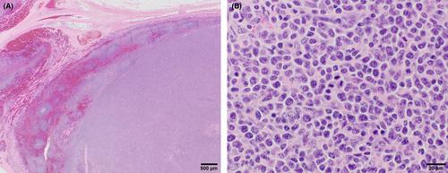
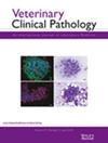
The patient was given a working diagnosis of enzootic bovine leukosis (EBL) and lymphoma based on CSF analysis and positive serum BLV ELISA (Cornell University Animal Health Diagnostic Center), which reports both high sensitivity and specificity.1 The owner elected euthanasia, given the persistent paresis despite flotation therapy and dexamethasone treatment. Postmortem histopathologic examination of multiple lymph nodes (retroperitoneal and cervical) was performed to support the antemortem diagnosis. The lymph node aspirate was critical to supporting the diagnostic findings of CSF analysis but was not performed as the initial diagnostic of choice due to concern that the result would not definitively diagnose EBL as the cause of the cow's paresis. The lymph nodes were multifocally to diffusely expanded by sheets of neoplastic round cells that distorted the nodal architecture. The cells had scant eosinophilic cytoplasm and large round nuclei approximately two to three times the diameter of an erythrocyte, with coarsely stippled chromatin and one to two prominent nucleoli. Nuclear pleomorphism was moderate, and mitotic figures numbered up to 22 per single high-power (2.37 mm2) field (Figure 2A,B).
Two clinical entities of lymphoma documented in cattle are sporadic and enzootic bovine leukosis, the latter being more common and found in association with bovine leukemia virus (BLV) infection.2, 3 BLV, a retrovirus, is known to be the causative agent of EBL and is a lymphoproliferative infection spread in secretions from infected cows.4 In this case, the farm had not previously tested for BLV, making the herd status unknown. BLV is reported to cause clinical syndromes in ≤5% of cattle, with more frequent reports in dairy cattle, specifically Holstein cows, and few reports in beef cattle breeds.3, 5 Common predilection sites include the spinal canal, heart, abomasum, uterus, and kidneys. Depending on the publication, up to 100% of cattle with reported spinal lymphoma also have lymph node involvement as identified on histopathologic biopsy, not solely on examination, as was identified in this case.6 In BLV-infected cattle, CBCs are often normal, with only 30% reportedly having a lymphocytosis.3
One causative etiology of recumbency in dairy cows is neoplasia. Diagnosis is often made on postmortem examination with the identification of an extradural mass. Of the reviews of extradural masses in cattle, neoplastic and non-neoplastic lesions have been diagnosed. Over 50% of all extradural masses and up to 78% of neoplastic extradural masses are reported to be lymphoma in a study of recumbent dairy cattle.7 CSF might be suggestive of a potential neoplastic process, but only rarely allows for diagnosis with lymphocytes often not seen on examination.5 Although CSF values commonly remain within the reference range, CSF protein and total nucleated cell counts are reported to be significantly increased in recumbent cattle with underlying neoplasia compared with other causes of recumbency.7 Nucleated cell counts are often reported to be increased, with the type of WBC seen on CSF cytology not being discussed. In one study, recumbent dairy cows had a median of 4.4 total nucleated cells /μL, while non-recumbent cows with spinal cord lesions had <1 total nucleated cells/μL.7 Thus, normal CSF does not rule out the possibility of lymphoma.
A diagnosis of EBL is multifaceted, with investigation of identified affected organ systems at the forefront to ensure an accurate diagnosis is made, with reinforcement from lymph node biopsy and BLV ELISA. BLV-associated syndromes are most noted in dairy breeds and commonly manifest with cardiac or gastrointestinal disease. Thus, a report of spinal lymphoma in an Angus cow is highly unusual. Spinal tumors are only identified in approximately 30% of cases of EBL.5 The antemortem diagnosis of lymphoma in this patient was made based on a combination of patient history and cytologic findings in the CSF, emphasizing the importance of CSF analysis in recumbent cows with a lack of response to standard treatment and exclusion of other common causes of recumbency, in conjunction with appropriate spinal fluid analysis.
期刊介绍:
Veterinary Clinical Pathology is the official journal of the American Society for Veterinary Clinical Pathology (ASVCP) and the European Society of Veterinary Clinical Pathology (ESVCP). The journal''s mission is to provide an international forum for communication and discussion of scientific investigations and new developments that advance the art and science of laboratory diagnosis in animals. Veterinary Clinical Pathology welcomes original experimental research and clinical contributions involving domestic, laboratory, avian, and wildlife species in the areas of hematology, hemostasis, immunopathology, clinical chemistry, cytopathology, surgical pathology, toxicology, endocrinology, laboratory and analytical techniques, instrumentation, quality assurance, and clinical pathology education.
 求助内容:
求助内容: 应助结果提醒方式:


