Spared ulnar nerve injury results in increased layer III–VI excitability in the pig somatosensory cortex
IF 5.9
3区 农林科学
Q1 VETERINARY SCIENCES
引用次数: 0
Abstract
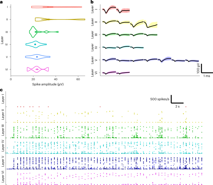
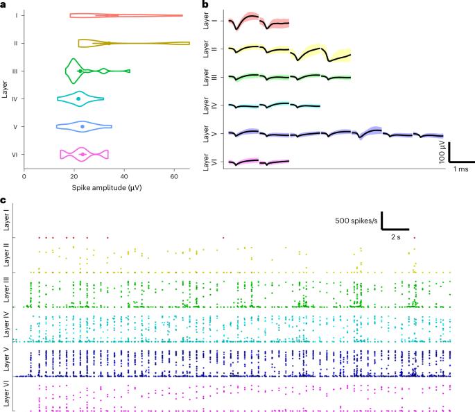
This study describes cortical recordings in a large animal nerve injury model. We investigated differences in primary somatosensory cortex (S1) hyperexcitability when stimulating injured and uninjured nerves and how different cortical layers contribute to S1 hyperexcitability after spared ulnar nerve injury. We used a multielectrode array to record single-neuron activity in the S1 of ten female Danish landrace pigs. Electrical stimulation of the injured and uninjured nerve evoked brain activity up to 3 h after injury. The peak amplitude and latency of early and late peristimulus time histogram responses were extracted for statistical analysis. Histological investigations determined the layer of the cortex in which each electrode contact was placed. Nerve injury increased the early peak amplitude compared with that of the control group. This difference was significant immediately after nerve injury when the uninjured nerve was stimulated, while it was delayed for the injured nerve. The amplitude of the early peak was increased in layers III–VI after nerve injury compared with the control. In layer III, S1 excitability was also increased compared with preinjury for the early peak. Furthermore, the late peak was significantly larger in layer III than in the other layers in the intervention and control group before and after injury. Thus, the most prominent increase in excitability occurred in layer III, which is responsible for the gain modulation of cortical output through layer V. Therefore, layer III neurons seem to have an important role in altered brain excitability after nerve injury. Meijs et al. perform an electrophysiological investigation of cortical responses in a pig nerve injury model, showing the role of layer III–VI neurons in altered primary somatosensory cortex excitability after nerve injury.


幸免的尺神经损伤会导致猪躯体感觉皮层 III-VI 层兴奋性增加。
本研究描述了大型动物神经损伤模型的皮层记录。我们研究了刺激受伤神经和未受伤神经时初级躯体感觉皮层(S1)过度兴奋性的差异,以及尺神经损伤后不同皮层对 S1 过度兴奋性的贡献。我们使用多电极阵列记录了 10 头雌性丹麦陆地猪 S1 的单神经元活动。电刺激受伤和未受伤的神经可诱发脑部活动,时间最长可达受伤后 3 小时。提取早期和晚期周围刺激时间直方图反应的峰值振幅和潜伏期进行统计分析。组织学调查确定了每个电极触点所在的皮层。与对照组相比,神经损伤增加了早期峰值振幅。神经损伤后,刺激未损伤神经时,这种差异立即显现,而刺激损伤神经时,这种差异则延迟显现。与对照组相比,神经损伤后第三至第六层的早期峰值振幅增大。在第三层,S1 兴奋性的早期峰值也比损伤前增加。此外,在干预组和对照组中,损伤前后第三层的晚峰值明显大于其他层。因此,第三层神经元在神经损伤后大脑兴奋性的改变中似乎起着重要作用。
本文章由计算机程序翻译,如有差异,请以英文原文为准。
求助全文
约1分钟内获得全文
求助全文
来源期刊

Lab Animal
农林科学-兽医学
CiteScore
0.60
自引率
2.90%
发文量
181
审稿时长
>36 weeks
期刊介绍:
LabAnimal is a Nature Research journal dedicated to in vivo science and technology that improves our basic understanding and use of model organisms of human health and disease. In addition to basic research, methods and technologies, LabAnimal also covers important news, business and regulatory matters that impact the development and application of model organisms for preclinical research.
LabAnimal's focus is on innovative in vivo methods, research and technology covering a wide range of model organisms. Our broad scope ensures that the work we publish reaches the widest possible audience. LabAnimal provides a rigorous and fair peer review of manuscripts, high standards for copyediting and production, and efficient publication.
 求助内容:
求助内容: 应助结果提醒方式:


