Fabiana Vitória Ananias Gonçalves, Orlando Aguirre Guedes, Sávio Akio Kachiyama, Larissa Pinzan Flauzino, Aurélio Rosa da Silva Júnior, Andreza Maria Fábio Aranha
{"title":"Assessment of tooth discoloration induced by root canal filling materials in pediatric dentistry.","authors":"Fabiana Vitória Ananias Gonçalves, Orlando Aguirre Guedes, Sávio Akio Kachiyama, Larissa Pinzan Flauzino, Aurélio Rosa da Silva Júnior, Andreza Maria Fábio Aranha","doi":"10.1590/0103-6440202405838","DOIUrl":null,"url":null,"abstract":"<p><p>This study investigated the potential for tooth discoloration of root canal filling pastes used in pediatric dentistry. Sixty bovine incisors were sectioned 2 mm apical to the cementoenamel junction and allocated into 6 groups (n = 10) according to the type of filling material used: G1- Zinc oxide-eugenol sealer; G2- Zinc oxide-eugenol and iodoform paste; G3- Calcium hydroxide (CH) and zinc oxide paste; G4- CH, zinc oxide, and iodoform paste; G5- CH and iodoform paste; and G6- Control. Polyethylene glycol 400 was used as a vehicle for CH-containing pastes. Color measurements were taken at specific intervals: preceding endodontic treatment (T0) and at successive points of 1 month (T1), 2 months (T2), 3 months (T3), and 1 year (T4) after the placement of the filling material. The color change (∆E) was calculated using the CIELab formula. Statistical analysis was performed using ANOVA, followed by Tukey's post hoc test (α = 5%). Significant differences were observed among the filling materials and time intervals (p <0.001). All groups exhibited color changes over time, except G1 and G5, which showed color changes only after 1 year. G1 and G2 demonstrated the highest ∆E values, with a statistically significant difference observed only at T2 when compared to G3 (p = 0.008). Root canal filling materials used in primary teeth have the potential to induce tooth discoloration.</p>","PeriodicalId":101363,"journal":{"name":"Brazilian dental journal","volume":"35 ","pages":"e245838"},"PeriodicalIF":0.0000,"publicationDate":"2024-09-16","publicationTypes":"Journal Article","fieldsOfStudy":null,"isOpenAccess":false,"openAccessPdf":"https://www.ncbi.nlm.nih.gov/pmc/articles/PMC11405006/pdf/","citationCount":"0","resultStr":null,"platform":"Semanticscholar","paperid":null,"PeriodicalName":"Brazilian dental journal","FirstCategoryId":"1085","ListUrlMain":"https://doi.org/10.1590/0103-6440202405838","RegionNum":0,"RegionCategory":null,"ArticlePicture":[],"TitleCN":null,"AbstractTextCN":null,"PMCID":null,"EPubDate":"2024/1/1 0:00:00","PubModel":"eCollection","JCR":"","JCRName":"","Score":null,"Total":0}
引用次数: 0
Abstract
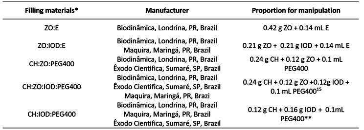
This study investigated the potential for tooth discoloration of root canal filling pastes used in pediatric dentistry. Sixty bovine incisors were sectioned 2 mm apical to the cementoenamel junction and allocated into 6 groups (n = 10) according to the type of filling material used: G1- Zinc oxide-eugenol sealer; G2- Zinc oxide-eugenol and iodoform paste; G3- Calcium hydroxide (CH) and zinc oxide paste; G4- CH, zinc oxide, and iodoform paste; G5- CH and iodoform paste; and G6- Control. Polyethylene glycol 400 was used as a vehicle for CH-containing pastes. Color measurements were taken at specific intervals: preceding endodontic treatment (T0) and at successive points of 1 month (T1), 2 months (T2), 3 months (T3), and 1 year (T4) after the placement of the filling material. The color change (∆E) was calculated using the CIELab formula. Statistical analysis was performed using ANOVA, followed by Tukey's post hoc test (α = 5%). Significant differences were observed among the filling materials and time intervals (p <0.001). All groups exhibited color changes over time, except G1 and G5, which showed color changes only after 1 year. G1 and G2 demonstrated the highest ∆E values, with a statistically significant difference observed only at T2 when compared to G3 (p = 0.008). Root canal filling materials used in primary teeth have the potential to induce tooth discoloration.

 求助内容:
求助内容: 应助结果提醒方式:


