Spatial proteomics and transcriptomics of the maternal-fetal interface in placenta accreta spectrum

IF 6.4 2区 医学 Q1 MEDICAL LABORATORY TECHNOLOGY
{"title":"Spatial proteomics and transcriptomics of the maternal-fetal interface in placenta accreta spectrum","authors":"","doi":"10.1016/j.trsl.2024.09.004","DOIUrl":null,"url":null,"abstract":"<div><div>In severe Placenta Accreta Spectrum (PAS), trophoblasts gain deep access in the myometrium (placenta increta). This study investigated alterations at the fetal-maternal interface in PAS cases using a systems biology approach consisting of immunohistochemistry, spatial transcriptomics and proteomics. We identified spatial variation in the distribution of CD4<sup>+</sup>, CD3<sup>+</sup> and CD8<sup>+</sup> T-cells at the maternal-interface in placenta increta cases. Spatial transcriptomics identified transcription factors involved in promotion of trophoblast invasion such as AP-1 subunits ATF-3 and JUN, and NFKB were upregulated in regions with deep myometrial invasion. Pathway analysis of differentially expressed genes demonstrated that degradation of extracellular matrix (ECM) and class 1 MHC protein were increased in increta regions, suggesting local tissue injury and immune suppression. Spatial proteomics demonstrated that increta regions were characterised by excessive trophoblastic proliferation in an immunosuppressive environment. Expression of inhibitors of apoptosis such as BCL-2 and fibronectin were increased, while CTLA-4 was decreased and increased expression of PD-L1, PD-L2 and CD14 macrophages. Additionally, CD44, which is a ligand of fibronectin that promotes trophoblast invasion and cell adhesion was also increased in increta regions. We subsequently examined ligand receptor interactions enriched in increta regions, with interactions with ITGβ1, including with fibronectin and ADAMS, emerging as central in increta. These ITGβ1 ligand interactions are involved in activation of epithelial–mesenchymal transition and remodelling of ECM suggesting a more invasive trophoblast phenotype. In PAS, we suggest this is driven by fibronectin via AP-1 signalling, likely as a secondary response to myometrial scarring.</div></div>","PeriodicalId":23226,"journal":{"name":"Translational Research","volume":null,"pages":null},"PeriodicalIF":6.4000,"publicationDate":"2024-09-28","publicationTypes":"Journal Article","fieldsOfStudy":null,"isOpenAccess":false,"openAccessPdf":"","citationCount":"0","resultStr":null,"platform":"Semanticscholar","paperid":null,"PeriodicalName":"Translational Research","FirstCategoryId":"3","ListUrlMain":"https://www.sciencedirect.com/science/article/pii/S1931524424001725","RegionNum":2,"RegionCategory":"医学","ArticlePicture":[],"TitleCN":null,"AbstractTextCN":null,"PMCID":null,"EPubDate":"","PubModel":"","JCR":"Q1","JCRName":"MEDICAL LABORATORY TECHNOLOGY","Score":null,"Total":0}
引用次数: 0

Abstract

In severe Placenta Accreta Spectrum (PAS), trophoblasts gain deep access in the myometrium (placenta increta). This study investigated alterations at the fetal-maternal interface in PAS cases using a systems biology approach consisting of immunohistochemistry, spatial transcriptomics and proteomics. We identified spatial variation in the distribution of CD4+, CD3+ and CD8+ T-cells at the maternal-interface in placenta increta cases. Spatial transcriptomics identified transcription factors involved in promotion of trophoblast invasion such as AP-1 subunits ATF-3 and JUN, and NFKB were upregulated in regions with deep myometrial invasion. Pathway analysis of differentially expressed genes demonstrated that degradation of extracellular matrix (ECM) and class 1 MHC protein were increased in increta regions, suggesting local tissue injury and immune suppression. Spatial proteomics demonstrated that increta regions were characterised by excessive trophoblastic proliferation in an immunosuppressive environment. Expression of inhibitors of apoptosis such as BCL-2 and fibronectin were increased, while CTLA-4 was decreased and increased expression of PD-L1, PD-L2 and CD14 macrophages. Additionally, CD44, which is a ligand of fibronectin that promotes trophoblast invasion and cell adhesion was also increased in increta regions. We subsequently examined ligand receptor interactions enriched in increta regions, with interactions with ITGβ1, including with fibronectin and ADAMS, emerging as central in increta. These ITGβ1 ligand interactions are involved in activation of epithelial–mesenchymal transition and remodelling of ECM suggesting a more invasive trophoblast phenotype. In PAS, we suggest this is driven by fibronectin via AP-1 signalling, likely as a secondary response to myometrial scarring.
胎盘早剥谱母胎界面的空间蛋白质组学和转录组学。
在严重的增厚性胎盘(PAS)中,滋养细胞深入子宫肌层(增厚性胎盘)。本研究采用免疫组化、空间转录组学和蛋白质组学组成的系统生物学方法,研究了PAS病例中胎儿与母体界面的变化。我们确定了增厚胎盘病例中母体界面 CD4+、CD3+ 和 CD8+ T 细胞分布的空间变化。空间转录组学发现了参与促进滋养细胞侵袭的转录因子,如AP-1亚基ATF-3和JUN,以及NFKB在子宫肌层侵袭区域上调。差异表达基因的通路分析表明,在增殖区细胞外基质(ECM)和1类MHC蛋白的降解增加,表明局部组织损伤和免疫抑制。空间蛋白质组学显示,增殖区的特点是滋养细胞在免疫抑制环境中过度增殖。凋亡抑制因子(如 BCL-2 和纤维粘连蛋白)的表达增加,而 CTLA-4 的表达减少,PD-L1、PD-L2 和 CD14 巨噬细胞的表达增加。此外,促进滋养细胞侵袭和细胞粘附的纤维粘连蛋白配体 CD44 在增殖区也有所增加。我们随后研究了在增殖区富集的配体受体相互作用,发现与 ITGβ1 的相互作用,包括与纤维粘连蛋白和 ADAMS 的相互作用,在增殖区非常重要。这些 ITGβ1 配体相互作用参与了上皮-间质转化的激活和 ECM 的重塑,这表明滋养层细胞表型更具侵袭性。在 PAS 中,我们认为这是由纤维连接蛋白通过 AP-1 信号驱动的,很可能是对子宫肌层瘢痕的次级反应。
本文章由计算机程序翻译,如有差异,请以英文原文为准。
求助全文
约1分钟内获得全文 求助全文
来源期刊
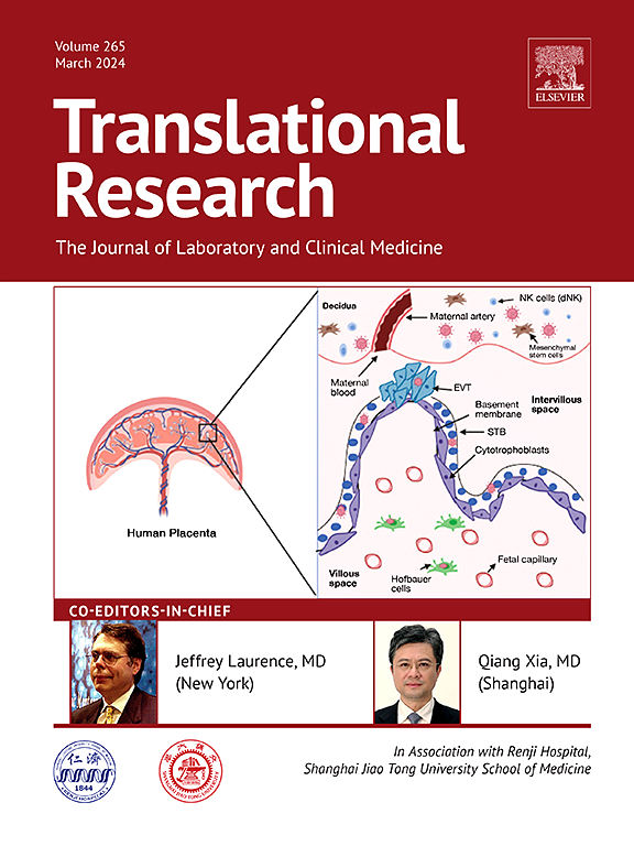
Translational Research
Translational Research 医学-医学:内科
CiteScore
15.70
自引率
0.00%
发文量
195
审稿时长
14 days
期刊介绍: Translational Research (formerly The Journal of Laboratory and Clinical Medicine) delivers original investigations in the broad fields of laboratory, clinical, and public health research. Published monthly since 1915, it keeps readers up-to-date on significant biomedical research from all subspecialties of medicine.
×
引用
GB/T 7714-2015
复制
MLA
复制
APA
复制
导出至
BibTeX EndNote RefMan NoteFirst NoteExpress
×
提示
您的信息不完整,为了账户安全,请先补充。
现在去补充
×
提示
您因"违规操作"
具体请查看互助需知
我知道了
×
提示
确定
请完成安全验证×
copy
已复制链接
快去分享给好友吧!
我知道了
右上角分享
点击右上角分享
0
联系我们:info@booksci.cn Book学术提供免费学术资源搜索服务,方便国内外学者检索中英文文献。致力于提供最便捷和优质的服务体验。 Copyright © 2023 布克学术 All rights reserved.
京ICP备2023020795号-1
ghs 京公网安备 11010802042870号
Book学术文献互助
Book学术文献互助群
群 号:481959085
Book学术官方微信