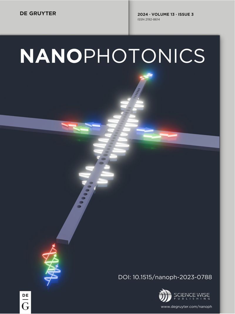
Image analysis optimization for nanowire-based optical detection of molecules
IF 6.5
2区 物理与天体物理
Q1 MATERIALS SCIENCE, MULTIDISCIPLINARY
引用次数: 0
Abstract
Semiconductor nanowires can enhance the signal of fluorescent molecules, thus significantly improving the limits of fluorescence detection in optical biosensing. In this work, we explore how the sensitivity can further be enhanced through “digital” detection of adequately spaced vertically aligned nanowires, employing single-emitter localization methods, and bright-field microscopy. Additionally, we introduce a systematic analysis pipeline aimed at harnessing this digital detection capability and evaluate its impact on detection sensitivity. Using a streptavidin-biotin assay, we demonstrate that single-emitter localization expands the dynamic range to encompass five orders of magnitude, enabling detections of concentrations ranging from 10 fM to 10 nM. This represents two to three orders of magnitude improvement in detection compared to methods that do not utilize single-emitter localization. We validate our analysis framework by simulating an artificial dataset based on numerical solutions of Maxwell’s equations. Furthermore, we benchmark our results against total internal reflection fluorescence microscopy and find, in time-resolved titration experiments, that nanowires offer higher sensitivity at the lowest concentrations, attributed to a combination of higher protein capture rate and higher intensity per single protein binding event. These findings suggest promising applications of nanowires in both endpoint and time-resolved biosensing.优化基于纳米线的分子光学检测图像分析
半导体纳米线可以增强荧光分子的信号,从而显著提高光学生物传感中荧光检测的极限。在这项工作中,我们探讨了如何通过 "数字 "检测垂直排列的适当间距纳米线、采用单发射极定位方法和明场显微镜来进一步提高灵敏度。此外,我们还介绍了旨在利用这种数字检测能力的系统分析管道,并评估了其对检测灵敏度的影响。利用链霉亲和素-生物素检测法,我们证明单发射极定位将动态范围扩大到了五个数量级,可检测的浓度范围从 10 fM 到 10 nM。与不使用单发射极定位的方法相比,这意味着检测能力提高了两到三个数量级。我们通过模拟基于麦克斯韦方程数值解的人工数据集来验证我们的分析框架。此外,我们还将我们的结果与全内反射荧光显微镜进行了比较,发现在时间分辨滴定实验中,纳米线在最低浓度下具有更高的灵敏度,这归因于更高的蛋白质捕获率和更高的单次蛋白质结合强度。这些发现表明,纳米线在终点和时间分辨生物传感中的应用前景广阔。
本文章由计算机程序翻译,如有差异,请以英文原文为准。
求助全文
约1分钟内获得全文
求助全文
来源期刊

Nanophotonics
NANOSCIENCE & NANOTECHNOLOGY-MATERIALS SCIENCE, MULTIDISCIPLINARY
CiteScore
13.50
自引率
6.70%
发文量
358
审稿时长
7 weeks
期刊介绍:
Nanophotonics, published in collaboration with Sciencewise, is a prestigious journal that showcases recent international research results, notable advancements in the field, and innovative applications. It is regarded as one of the leading publications in the realm of nanophotonics and encompasses a range of article types including research articles, selectively invited reviews, letters, and perspectives.
The journal specifically delves into the study of photon interaction with nano-structures, such as carbon nano-tubes, nano metal particles, nano crystals, semiconductor nano dots, photonic crystals, tissue, and DNA. It offers comprehensive coverage of the most up-to-date discoveries, making it an essential resource for physicists, engineers, and material scientists.
 求助内容:
求助内容: 应助结果提醒方式:


