In vitro vascularization of 3D cell aggregates in microwells with integrated vascular beds
IF 8.7
1区 医学
Q1 ENGINEERING, BIOMEDICAL
引用次数: 0
Abstract
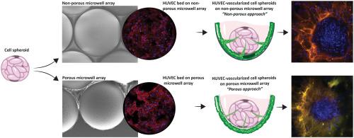
Most human tissues possess vascular networks supplying oxygen and nutrients. Engineering of functional tissue and organ models or equivalents often require the integration of artificial vascular networks. Several approaches, such as organs on chips and three-dimensional (3D) bioprinting, have been pursued to obtain vasculature and vascularized tissues in vitro. This technical feasibility study proposes a new approach for the in vitro vascularization of 3D microtissues. For this, we thermoform arrays of round-bottom microwells into thin non-porous and porous polymer films/membranes and culture vascular beds on them from which endothelial sprouting occurs in a Matrigel-based 3D extra cellular matrix. We present two possible culture configurations for the microwell-integrated vascular beds. In the first configuration, human umbilical vein endothelial cells (HUVECs) grow on and sprout from the inner wall of the non-porous microwells. In the second one, HUVECs grow on the outer surface of the porous microwells and sprout through the pores toward the inside. These approaches are extended to lymphatic endothelial cells. As a proof of concept, we demonstrate the in vitro vascularization of spheroids from human mesenchymal stem cells and MG-63 human osteosarcoma cells. Our results show the potential of this approach to provide the spheroids with an abundant outer vascular network and the indication of an inner vasculature.
集成血管床的微孔中三维细胞聚集体的体外血管化
大多数人体组织都拥有供应氧气和营养物质的血管网络。功能性组织和器官模型或等效模型的工程设计往往需要整合人工血管网络。目前已有多种方法,如芯片上的器官和三维(3D)生物打印,用于获取体外血管和血管化组织。这项技术可行性研究提出了一种三维微组织体外血管化的新方法。为此,我们将圆底微孔阵列热成型为薄的无孔和多孔聚合物薄膜/膜,并在其上培养血管床,在基于 Matrigel 的三维外细胞基质中,内皮从中萌发。我们介绍了微孔集成血管床的两种可能培养配置。在第一种配置中,人脐静脉内皮细胞(HUVEC)生长在无孔微孔的内壁上并从内壁发芽。在第二种配置中,人脐静脉内皮细胞(HUVEC)生长在多孔微孔的外表面,并通过孔隙向内发芽。这些方法已扩展到淋巴内皮细胞。作为概念验证,我们展示了人类间充质干细胞和 MG-63 人类骨肉瘤细胞球体的体外血管化。我们的研究结果表明,这种方法有可能为球体提供丰富的外层血管网络,并显示出内层血管。
本文章由计算机程序翻译,如有差异,请以英文原文为准。
求助全文
约1分钟内获得全文
求助全文
来源期刊
Materials Today Bio
Multiple-
CiteScore
8.30
自引率
4.90%
发文量
303
审稿时长
30 days
期刊介绍:
Materials Today Bio is a multidisciplinary journal that specializes in the intersection between biology and materials science, chemistry, physics, engineering, and medicine. It covers various aspects such as the design and assembly of new structures, their interaction with biological systems, functionalization, bioimaging, therapies, and diagnostics in healthcare. The journal aims to showcase the most significant advancements and discoveries in this field. As part of the Materials Today family, Materials Today Bio provides rigorous peer review, quick decision-making, and high visibility for authors. It is indexed in Scopus, PubMed Central, Emerging Sources, Citation Index (ESCI), and Directory of Open Access Journals (DOAJ).
 求助内容:
求助内容: 应助结果提醒方式:


