Activation of Piezo1 by intracranial hypertension induced neuronal apoptosis via activating hippo pathway

IF 4.8 1区 医学 Q1 NEUROSCIENCES
Jia Zeng, Zhen Fang, Jiajia Duan, Zichen Zhang, Yunzhi Wang, Yiping Wang, Lei Chen, Jikai Wang, Fei Liu
{"title":"Activation of Piezo1 by intracranial hypertension induced neuronal apoptosis via activating hippo pathway","authors":"Jia Zeng,&nbsp;Zhen Fang,&nbsp;Jiajia Duan,&nbsp;Zichen Zhang,&nbsp;Yunzhi Wang,&nbsp;Yiping Wang,&nbsp;Lei Chen,&nbsp;Jikai Wang,&nbsp;Fei Liu","doi":"10.1111/cns.14872","DOIUrl":null,"url":null,"abstract":"<div>\n \n \n <section>\n \n <h3> Aim</h3>\n \n <p>Most of the subarachnoid hemorrhage (SAH) patients experienced the symptom of severe headache caused by intracranial hypertension. Piezo1 is a mechanosensitive ion channel protein. This study aimed to investigate the effect of Piezo1 on neurons in response to intracranial hypertension.</p>\n </section>\n \n <section>\n \n <h3> Methods</h3>\n \n <p>The SAH rat model was performed by the modified endovascular perforation method. Piezo1 inhibitor GsMTx4 was administered intraperitoneally after SAH induction. To investigate the underlying mechanism, the selective Piezo1 agonist Yoda1, Piezo1 shRNA, and MY-875 were administered via intracerebroventricular injection before SAH induction. In vitro, we designed a pressurizing device to exclusively explore the effect of Piezo1 activation on primary neurons. Neurons were pretreated with Piezo1 inhibition followed by intracranial hypertension treatment, and then apoptosis-related proteins were detected.</p>\n </section>\n \n <section>\n \n <h3> Results</h3>\n \n <p>Piezo1 inhibition significantly attenuated neuronal apoptosis and improved the outcome of neurological deficits in rats after SAH. The Hippo pathway agonist MY-875 reversed the anti-apoptotic effects of Piezo1 knockdown. In vitro, intracranial hypertension mimicked by the pressurizing device induced Piezo1 expression, resulting in Hippo pathway activation and neuronal apoptosis. The Hippo pathway inhibitor Xmu-mp-1 attenuated Yoda1-induced neuronal apoptosis. In addition, the combination of hypertension and oxyhemoglobin treatment exacerbated neuronal apoptosis.</p>\n </section>\n \n <section>\n \n <h3> Conclusions</h3>\n \n <p>Intracranial hypertension induced Piezo1 expression, neuronal apoptosis, and the Hippo pathway activation; the Hippo signaling pathway is involved in Piezo1 activation-induced neuronal apoptosis in respond to intracranial hypertension. Primary neurons treated with intracranial hypertension and oxyhemoglobin together can better characterize the circumstance of SAH in vivo, which is contributed to construct an ideal in vitro SAH model.</p>\n </section>\n </div>","PeriodicalId":154,"journal":{"name":"CNS Neuroscience & Therapeutics","volume":"30 9","pages":""},"PeriodicalIF":4.8000,"publicationDate":"2024-09-27","publicationTypes":"Journal Article","fieldsOfStudy":null,"isOpenAccess":false,"openAccessPdf":"https://onlinelibrary.wiley.com/doi/epdf/10.1111/cns.14872","citationCount":"0","resultStr":null,"platform":"Semanticscholar","paperid":null,"PeriodicalName":"CNS Neuroscience & Therapeutics","FirstCategoryId":"3","ListUrlMain":"https://onlinelibrary.wiley.com/doi/10.1111/cns.14872","RegionNum":1,"RegionCategory":"医学","ArticlePicture":[],"TitleCN":null,"AbstractTextCN":null,"PMCID":null,"EPubDate":"","PubModel":"","JCR":"Q1","JCRName":"NEUROSCIENCES","Score":null,"Total":0}
引用次数: 0

Abstract

Aim

Most of the subarachnoid hemorrhage (SAH) patients experienced the symptom of severe headache caused by intracranial hypertension. Piezo1 is a mechanosensitive ion channel protein. This study aimed to investigate the effect of Piezo1 on neurons in response to intracranial hypertension.

Methods

The SAH rat model was performed by the modified endovascular perforation method. Piezo1 inhibitor GsMTx4 was administered intraperitoneally after SAH induction. To investigate the underlying mechanism, the selective Piezo1 agonist Yoda1, Piezo1 shRNA, and MY-875 were administered via intracerebroventricular injection before SAH induction. In vitro, we designed a pressurizing device to exclusively explore the effect of Piezo1 activation on primary neurons. Neurons were pretreated with Piezo1 inhibition followed by intracranial hypertension treatment, and then apoptosis-related proteins were detected.

Results

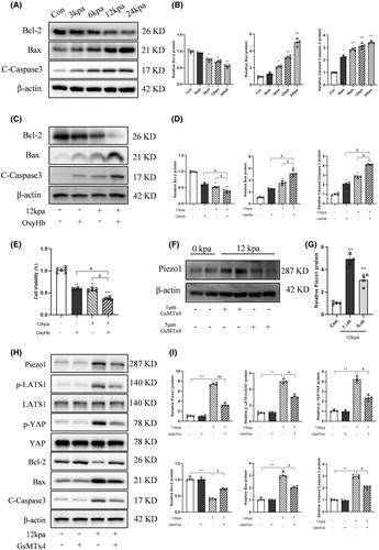
Piezo1 inhibition significantly attenuated neuronal apoptosis and improved the outcome of neurological deficits in rats after SAH. The Hippo pathway agonist MY-875 reversed the anti-apoptotic effects of Piezo1 knockdown. In vitro, intracranial hypertension mimicked by the pressurizing device induced Piezo1 expression, resulting in Hippo pathway activation and neuronal apoptosis. The Hippo pathway inhibitor Xmu-mp-1 attenuated Yoda1-induced neuronal apoptosis. In addition, the combination of hypertension and oxyhemoglobin treatment exacerbated neuronal apoptosis.

Conclusions

Intracranial hypertension induced Piezo1 expression, neuronal apoptosis, and the Hippo pathway activation; the Hippo signaling pathway is involved in Piezo1 activation-induced neuronal apoptosis in respond to intracranial hypertension. Primary neurons treated with intracranial hypertension and oxyhemoglobin together can better characterize the circumstance of SAH in vivo, which is contributed to construct an ideal in vitro SAH model.
Abstract Image

颅内高压激活 Piezo1,通过激活 hippo 通路诱导神经细胞凋亡
目的 大多数蛛网膜下腔出血(SAH)患者都有颅内高压引起的剧烈头痛症状。Piezo1 是一种机械敏感性离子通道蛋白。本研究旨在探讨 Piezo1 在颅内高压反应中对神经元的影响。 方法 SAH 大鼠模型采用改良血管内穿孔法。诱导 SAH 后腹腔注射 Piezo1 抑制剂 GsMTx4。为了研究其潜在机制,我们在诱导 SAH 之前通过脑室内注射选择性 Piezo1 激动剂 Yoda1、Piezo1 shRNA 和 MY-875。在体外,我们设计了一个加压装置,专门探讨 Piezo1 激活对初级神经元的影响。先对神经元进行 Piezo1 抑制预处理,然后进行颅内高压处理,最后检测凋亡相关蛋白。 结果 Piezo1 抑制剂能明显减轻 SAH 后大鼠神经元凋亡,改善神经功能缺损的预后。Hippo 通路激动剂 MY-875 逆转了 Piezo1 敲除的抗凋亡作用。在体外,加压装置模拟的颅内高压诱导了Piezo1的表达,导致Hippo通路激活和神经元凋亡。Hippo通路抑制剂Xmu-mp-1可减轻Yoda1诱导的神经细胞凋亡。此外,高血压和氧合血红蛋白联合治疗会加剧神经元凋亡。 结论 颅内高血压诱导 Piezo1 表达、神经元凋亡和 Hippo 通路激活;Hippo 信号通路参与了 Piezo1 激活诱导神经元凋亡对颅内高血压的反应。用颅内高压和氧合血红蛋白处理原代神经元能更好地描述 SAH 在体内的情况,有助于构建理想的体外 SAH 模型。
本文章由计算机程序翻译,如有差异,请以英文原文为准。
求助全文
约1分钟内获得全文 求助全文
来源期刊
CNS Neuroscience & Therapeutics
CNS Neuroscience & Therapeutics 医学-神经科学
CiteScore
7.30
自引率
12.70%
发文量
240
审稿时长
2 months
期刊介绍: CNS Neuroscience & Therapeutics provides a medium for rapid publication of original clinical, experimental, and translational research papers, timely reviews and reports of novel findings of therapeutic relevance to the central nervous system, as well as papers related to clinical pharmacology, drug development and novel methodologies for drug evaluation. The journal focuses on neurological and psychiatric diseases such as stroke, Parkinson’s disease, Alzheimer’s disease, depression, schizophrenia, epilepsy, and drug abuse.
×
引用
GB/T 7714-2015
复制
MLA
复制
APA
复制
导出至
BibTeX EndNote RefMan NoteFirst NoteExpress
×
提示
您的信息不完整,为了账户安全,请先补充。
现在去补充
×
提示
您因"违规操作"
具体请查看互助需知
我知道了
×
提示
确定
请完成安全验证×
copy
已复制链接
快去分享给好友吧!
我知道了
右上角分享
点击右上角分享
0
联系我们:info@booksci.cn Book学术提供免费学术资源搜索服务,方便国内外学者检索中英文文献。致力于提供最便捷和优质的服务体验。 Copyright © 2023 布克学术 All rights reserved.
京ICP备2023020795号-1
ghs 京公网安备 11010802042870号
Book学术文献互助
Book学术文献互助群
群 号:481959085
Book学术官方微信