Structural characterization of hierarchical polymer foams by combining X-ray micro-computed tomography and scanning electron microscopy
IF 5
2区 材料科学
Q1 MATERIALS SCIENCE, CHARACTERIZATION & TESTING
引用次数: 0
Abstract
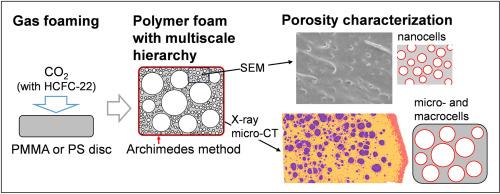
Complementary structural characterization methods are useful for studying the hierarchical cellular morphology of polymer foams. In this study, we employed scanning electron microscopy (SEM) and X-ray micro-computed tomography (micro-CT) to characterize the hierarchical cellular morphology of poly(methyl methacrylate) (PMMA) and polystyrene (PS) foams. The polymer foams were prepared using pure CO2 gas and CO2–chlorodifluoromethane (HCFC-22) gas mixtures as blowing agents. Depending on the type of polymer and HCFC-22 concentration, hierarchical cellular structures consisting of nanocells, microcells, and macrocells were obtained. The size distribution of the nanocells was determined by high-magnification SEM, while the size, shape, and spatial distribution of the microcells and macrocells in three dimensions were determined by micro-CT. Moreover, a well-designed micro-CT experiment enabled a brightness comparison between the foams and relative local density mapping of the foams based on the brightness. The results clearly showed the formation of a dense skin layer at the air interface of both PMMA and PS foams and dense matrix around the large macrocells in the PMMA foams. Thus, combining SEM and micro-CT provides a deeper understanding of the formation mechanism of the hierarchical cellular structure of polymer foams.

结合 X 射线微型计算机断层扫描和扫描电子显微镜分析分层聚合物泡沫的结构特征
互补的结构表征方法有助于研究聚合物泡沫的分层细胞形态。在这项研究中,我们采用扫描电子显微镜(SEM)和 X 射线显微计算机断层扫描(micro-CT)表征了聚(甲基丙烯酸甲酯)(PMMA)和聚苯乙烯(PS)泡沫的分层细胞形态。聚合物泡沫是使用纯二氧化碳气体和二氧化碳-氯二氟甲烷(HCFC-22)气体混合物作为发泡剂制备的。根据聚合物类型和 HCFC-22 浓度的不同,获得了由纳米细胞、微细胞和大细胞组成的分层细胞结构。高倍扫描电子显微镜测定了纳米细胞的尺寸分布,而显微 CT 则测定了微细胞和大细胞的尺寸、形状和三维空间分布。此外,通过精心设计的显微 CT 实验,还可以比较泡沫的亮度,并根据亮度绘制泡沫的相对局部密度图。结果清楚地表明,在 PMMA 和 PS 泡沫的空气界面上都形成了致密的表皮层,而在 PMMA 泡沫中,大型大孔周围则形成了致密的基质。因此,结合 SEM 和 micro-CT 可以更深入地了解聚合物泡沫分层细胞结构的形成机制。
本文章由计算机程序翻译,如有差异,请以英文原文为准。
求助全文
约1分钟内获得全文
求助全文
来源期刊

Polymer Testing
工程技术-材料科学:表征与测试
CiteScore
10.70
自引率
5.90%
发文量
328
审稿时长
44 days
期刊介绍:
Polymer Testing focuses on the testing, analysis and characterization of polymer materials, including both synthetic and natural or biobased polymers. Novel testing methods and the testing of novel polymeric materials in bulk, solution and dispersion is covered. In addition, we welcome the submission of the testing of polymeric materials for a wide range of applications and industrial products as well as nanoscale characterization.
The scope includes but is not limited to the following main topics:
Novel testing methods and Chemical analysis
• mechanical, thermal, electrical, chemical, imaging, spectroscopy, scattering and rheology
Physical properties and behaviour of novel polymer systems
• nanoscale properties, morphology, transport properties
Degradation and recycling of polymeric materials when combined with novel testing or characterization methods
• degradation, biodegradation, ageing and fire retardancy
Modelling and Simulation work will be only considered when it is linked to new or previously published experimental results.
 求助内容:
求助内容: 应助结果提醒方式:


