Human performance in predicting enhancement quality of gliomas using gadolinium-free MRI sequences

IF 2.3 4区 医学 Q3 CLINICAL NEUROLOGY
Aynur Azizova, Ivar J. H. G. Wamelink, Yeva Prysiazhniuk, Marcus Cakmak, Elif Kaya, Jan Petr, Frederik Barkhof, Vera C. Keil
{"title":"Human performance in predicting enhancement quality of gliomas using gadolinium-free MRI sequences","authors":"Aynur Azizova,&nbsp;Ivar J. H. G. Wamelink,&nbsp;Yeva Prysiazhniuk,&nbsp;Marcus Cakmak,&nbsp;Elif Kaya,&nbsp;Jan Petr,&nbsp;Frederik Barkhof,&nbsp;Vera C. Keil","doi":"10.1111/jon.13233","DOIUrl":null,"url":null,"abstract":"<div>\n \n \n <section>\n \n <h3> Background and Purpose</h3>\n \n <p>To develop and test a decision tree for predicting contrast enhancement quality and shape using precontrast magnetic resonance imaging (MRI) sequences in a large adult-type diffuse glioma cohort.</p>\n </section>\n \n <section>\n \n <h3> Methods</h3>\n \n <p>Preoperative MRI scans (development/optimization/test sets: <i>n</i> = 31/38/303, male = 17/22/189, mean age = 52/59/56.7 years, high-grade glioma = 22/33/249) were retrospectively evaluated, including pre- and postcontrast T1-weighted, T2-weighted, fluid-attenuated inversion recovery, and diffusion-weighted imaging sequences. Enhancement prediction decision tree (EPDT) was developed using development and optimization sets, incorporating four imaging features: necrosis, diffusion restriction, T2 inhomogeneity, and nonenhancing tumor margins. EPDT accuracy was assessed on a test set by three raters of variable experience. True enhancement features (gold standard) were evaluated using pre- and postcontrast T1-weighted images. Statistical analysis used confusion matrices, Cohen's/Fleiss’ kappa, and Kendall's <i>W</i>. Significance threshold was <i>p</i> &lt; .05.</p>\n </section>\n \n <section>\n \n <h3> Results</h3>\n \n <p>Raters 1, 2, and 3 achieved overall accuracies of .86 (95% confidence interval [CI]: .81-.90), .89 (95% CI: .85-.92), and .92 (95% CI: .89-.95), respectively, in predicting enhancement quality (marked, mild, or no enhancement). Regarding shape, defined as the thickness of enhancing margin (solid, rim, or no enhancement), accuracies were .84 (95% CI: .79-.88), .88 (95% CI: .84-.92), and .89 (95% CI: .85-.92). Intrarater intergroup agreement comparing predicted and true enhancement features consistently reached substantial levels (≥.68 [95% CI: .61-.75]). Interrater comparison showed at least moderate agreement (group: ≥.42 [95% CI: .36-.48], pairwise: ≥.61 [95% CI: .50-.72]). Among the imaging features in the EPDT, necrosis assessment displayed the highest intra- and interrater consistency (≥.80 [95% CI: .73-.88]).</p>\n </section>\n \n <section>\n \n <h3> Conclusion</h3>\n \n <p>The proposed EPDT has high accuracy in predicting enhancement patterns of gliomas irrespective of rater experience.</p>\n </section>\n </div>","PeriodicalId":16399,"journal":{"name":"Journal of Neuroimaging","volume":"34 6","pages":"673-693"},"PeriodicalIF":2.3000,"publicationDate":"2024-09-19","publicationTypes":"Journal Article","fieldsOfStudy":null,"isOpenAccess":false,"openAccessPdf":"https://onlinelibrary.wiley.com/doi/epdf/10.1111/jon.13233","citationCount":"0","resultStr":null,"platform":"Semanticscholar","paperid":null,"PeriodicalName":"Journal of Neuroimaging","FirstCategoryId":"3","ListUrlMain":"https://onlinelibrary.wiley.com/doi/10.1111/jon.13233","RegionNum":4,"RegionCategory":"医学","ArticlePicture":[],"TitleCN":null,"AbstractTextCN":null,"PMCID":null,"EPubDate":"","PubModel":"","JCR":"Q3","JCRName":"CLINICAL NEUROLOGY","Score":null,"Total":0}
引用次数: 0

Abstract

Background and Purpose

To develop and test a decision tree for predicting contrast enhancement quality and shape using precontrast magnetic resonance imaging (MRI) sequences in a large adult-type diffuse glioma cohort.

Methods

Preoperative MRI scans (development/optimization/test sets: n = 31/38/303, male = 17/22/189, mean age = 52/59/56.7 years, high-grade glioma = 22/33/249) were retrospectively evaluated, including pre- and postcontrast T1-weighted, T2-weighted, fluid-attenuated inversion recovery, and diffusion-weighted imaging sequences. Enhancement prediction decision tree (EPDT) was developed using development and optimization sets, incorporating four imaging features: necrosis, diffusion restriction, T2 inhomogeneity, and nonenhancing tumor margins. EPDT accuracy was assessed on a test set by three raters of variable experience. True enhancement features (gold standard) were evaluated using pre- and postcontrast T1-weighted images. Statistical analysis used confusion matrices, Cohen's/Fleiss’ kappa, and Kendall's W. Significance threshold was p < .05.

Results

Raters 1, 2, and 3 achieved overall accuracies of .86 (95% confidence interval [CI]: .81-.90), .89 (95% CI: .85-.92), and .92 (95% CI: .89-.95), respectively, in predicting enhancement quality (marked, mild, or no enhancement). Regarding shape, defined as the thickness of enhancing margin (solid, rim, or no enhancement), accuracies were .84 (95% CI: .79-.88), .88 (95% CI: .84-.92), and .89 (95% CI: .85-.92). Intrarater intergroup agreement comparing predicted and true enhancement features consistently reached substantial levels (≥.68 [95% CI: .61-.75]). Interrater comparison showed at least moderate agreement (group: ≥.42 [95% CI: .36-.48], pairwise: ≥.61 [95% CI: .50-.72]). Among the imaging features in the EPDT, necrosis assessment displayed the highest intra- and interrater consistency (≥.80 [95% CI: .73-.88]).

Conclusion

The proposed EPDT has high accuracy in predicting enhancement patterns of gliomas irrespective of rater experience.
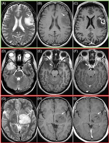
Abstract Image

利用无钆磁共振成像序列预测胶质瘤增强质量的人类表现。
背景与目的:在一个大型成人型弥漫性胶质瘤队列中开发并测试一种决策树,用于利用对比前磁共振成像(MRI)序列预测对比增强的质量和形状:对术前 MRI 扫描(开发/优化/测试集:n = 31/38/303,男性 = 17/22/189,平均年龄 = 52/59/56.7岁,高级别胶质瘤 = 22/33/249)进行回顾性评估,包括对比前和对比后 T1 加权、T2 加权、流体增强反转恢复和弥散加权成像序列。利用开发和优化集开发了增强预测决策树(EPDT),其中包含四个成像特征:坏死、弥散受限、T2 不均匀性和肿瘤边缘不增强。EPDT 的准确性由三位经验各异的评定者在测试集上进行评估。使用对比前和对比后的 T1 加权图像对真实增强特征(金标准)进行评估。统计分析采用混淆矩阵、Cohen's/Fleiss' kappa 和 Kendall's W:评分者 1、2 和 3 预测增强质量(明显、轻度或无增强)的总体准确率分别为 0.86(95% 置信区间 [CI]:0.81-.90)、0.89(95% CI:0.85-.92)和 0.92(95% CI:0.89-.95)。关于形状,即增强边缘的厚度(实心、边缘或无增强),准确率分别为 0.84(95% CI:0.79-.88)、0.88(95% CI:0.84-.92)和 0.89(95% CI:0.85-.92)。比较预测增强特征和真实增强特征的组间一致性达到了相当高的水平(≥.68 [95% CI: .61-.75])。相互比较至少显示出中等程度的一致性(组间:≥.42 [95% CI:.36-.48],配对:≥.61 [95% CI:.50-.72])。在 EPDT 的影像学特征中,坏死评估显示出最高的内部和相互间一致性(≥.80 [95% CI:.73-.88]):无论评分者的经验如何,所提出的 EPDT 在预测胶质瘤的增强模式方面都具有很高的准确性。
本文章由计算机程序翻译,如有差异,请以英文原文为准。
求助全文
约1分钟内获得全文 求助全文
来源期刊
Journal of Neuroimaging
Journal of Neuroimaging 医学-核医学
CiteScore
4.70
自引率
0.00%
发文量
117
审稿时长
6-12 weeks
期刊介绍: Start reading the Journal of Neuroimaging to learn the latest neurological imaging techniques. The peer-reviewed research is written in a practical clinical context, giving you the information you need on: MRI CT Carotid Ultrasound and TCD SPECT PET Endovascular Surgical Neuroradiology Functional MRI Xenon CT and other new and upcoming neuroscientific modalities.The Journal of Neuroimaging addresses the full spectrum of human nervous system disease, including stroke, neoplasia, degenerating and demyelinating disease, epilepsy, tumors, lesions, infectious disease, cerebral vascular arterial diseases, toxic-metabolic disease, psychoses, dementias, heredo-familial disease, and trauma.Offering original research, review articles, case reports, neuroimaging CPCs, and evaluations of instruments and technology relevant to the nervous system, the Journal of Neuroimaging focuses on useful clinical developments and applications, tested techniques and interpretations, patient care, diagnostics, and therapeutics. Start reading today!
×
引用
GB/T 7714-2015
复制
MLA
复制
APA
复制
导出至
BibTeX EndNote RefMan NoteFirst NoteExpress
×
提示
您的信息不完整,为了账户安全,请先补充。
现在去补充
×
提示
您因"违规操作"
具体请查看互助需知
我知道了
×
提示
确定
请完成安全验证×
copy
已复制链接
快去分享给好友吧!
我知道了
右上角分享
点击右上角分享
0
联系我们:info@booksci.cn Book学术提供免费学术资源搜索服务,方便国内外学者检索中英文文献。致力于提供最便捷和优质的服务体验。 Copyright © 2023 布克学术 All rights reserved.
京ICP备2023020795号-1
ghs 京公网安备 11010802042870号
Book学术文献互助
Book学术文献互助群
群 号:481959085
Book学术官方微信