Yun Zhao, Jiagen Li, Zhongkun Ji, Shasha Yu, Jinyong Lin, Hong Zhao
{"title":"Clinicopathological Features and Management of Orbital Cholesterol Granuloma.","authors":"Yun Zhao, Jiagen Li, Zhongkun Ji, Shasha Yu, Jinyong Lin, Hong Zhao","doi":"10.4103/joco.joco_200_23","DOIUrl":null,"url":null,"abstract":"<p><strong>Purpose: </strong>To investigate the clinical features, radiographic features, treatment strategies, pathological features, and prognosis of orbital cholesterol granuloma (CG).</p><p><strong>Methods: </strong>Twelve patients with orbital CG who were referred to Tianjin Eye Hospital between January 2002 and December 2020 were included in this retrospective case series study. Data collected including patient ophthalmic manifestations, imaging findings, treatment strategies, pathological features, and prognosis were retrospectively reviewed.</p><p><strong>Results: </strong>The patients comprised 10 males and 2 females. The mean age was 34.5 years (standard deviation [SD] = 8.9, median: 36 and range: 16-45 years). Four patients had a history of orbital trauma. The clinical manifestations at the first visit were proptosis (7/12, 58.3%), periorbital or eyelid swelling (6/12, 50%), limitation of eye movement (4/12, 33.3%), ptosis (2/12, 16.7%), and decreased visual acuity (1/12, 8.3%). Computed tomography (CT) showed a nonenhancing, well-circumscribed lesion in the orbit with extensive erosion of the adjacent frontal bone and temporal bone. Magnetic resonance imaging (MRI) showed a nonenhancing mass with intermediate-to-high signal intensity on T1- and T2-weighted images. Ten patients underwent lateral orbitotomy, and two patients underwent supraorbital orbitotomy. All patients had aggressive bone erosion. Histopathologic evaluation of the cyst contents and wall revealed cholesterol clefts, multinucleated giant cells, histiocytes, foamy macrophages, and altered blood pigments. The mean follow-up time of 79.6 months (SD = 49.8, range: 19-193 months). Three patients were lost to follow-up. No postoperative diminution of vision was noted, and no recurrence was observed.</p><p><strong>Conclusions: </strong>CGs can present as superotemporal or temporal orbital lesions. The diagnosis can be established based on CT and MRI. Most of the patients can have no history of orbital trauma.</p>","PeriodicalId":15423,"journal":{"name":"Journal of Current Ophthalmology","volume":"35 4","pages":"401-404"},"PeriodicalIF":0.9000,"publicationDate":"2024-08-10","publicationTypes":"Journal Article","fieldsOfStudy":null,"isOpenAccess":false,"openAccessPdf":"https://www.ncbi.nlm.nih.gov/pmc/articles/PMC11392298/pdf/","citationCount":"0","resultStr":null,"platform":"Semanticscholar","paperid":null,"PeriodicalName":"Journal of Current Ophthalmology","FirstCategoryId":"1085","ListUrlMain":"https://doi.org/10.4103/joco.joco_200_23","RegionNum":0,"RegionCategory":null,"ArticlePicture":[],"TitleCN":null,"AbstractTextCN":null,"PMCID":null,"EPubDate":"2023/10/1 0:00:00","PubModel":"eCollection","JCR":"Q3","JCRName":"OPHTHALMOLOGY","Score":null,"Total":0}
引用次数: 0
Abstract
Purpose: To investigate the clinical features, radiographic features, treatment strategies, pathological features, and prognosis of orbital cholesterol granuloma (CG).
Methods: Twelve patients with orbital CG who were referred to Tianjin Eye Hospital between January 2002 and December 2020 were included in this retrospective case series study. Data collected including patient ophthalmic manifestations, imaging findings, treatment strategies, pathological features, and prognosis were retrospectively reviewed.
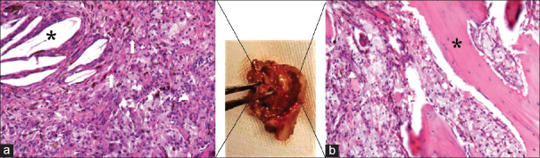
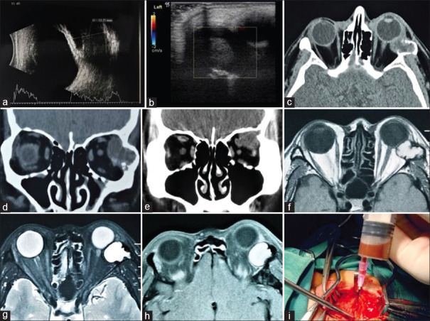
Results: The patients comprised 10 males and 2 females. The mean age was 34.5 years (standard deviation [SD] = 8.9, median: 36 and range: 16-45 years). Four patients had a history of orbital trauma. The clinical manifestations at the first visit were proptosis (7/12, 58.3%), periorbital or eyelid swelling (6/12, 50%), limitation of eye movement (4/12, 33.3%), ptosis (2/12, 16.7%), and decreased visual acuity (1/12, 8.3%). Computed tomography (CT) showed a nonenhancing, well-circumscribed lesion in the orbit with extensive erosion of the adjacent frontal bone and temporal bone. Magnetic resonance imaging (MRI) showed a nonenhancing mass with intermediate-to-high signal intensity on T1- and T2-weighted images. Ten patients underwent lateral orbitotomy, and two patients underwent supraorbital orbitotomy. All patients had aggressive bone erosion. Histopathologic evaluation of the cyst contents and wall revealed cholesterol clefts, multinucleated giant cells, histiocytes, foamy macrophages, and altered blood pigments. The mean follow-up time of 79.6 months (SD = 49.8, range: 19-193 months). Three patients were lost to follow-up. No postoperative diminution of vision was noted, and no recurrence was observed.
Conclusions: CGs can present as superotemporal or temporal orbital lesions. The diagnosis can be established based on CT and MRI. Most of the patients can have no history of orbital trauma.
期刊介绍:
Peer Review under the responsibility of Iranian Society of Ophthalmology Journal of Current Ophthalmology, the official publication of the Iranian Society of Ophthalmology, is a peer-reviewed, open-access, scientific journal that welcomes high quality original articles related to vision science and all fields of ophthalmology. Journal of Current Ophthalmology is the continuum of Iranian Journal of Ophthalmology published since 1969.

 求助内容:
求助内容: 应助结果提醒方式:


