Perturbed cell cycle phase-dependent positioning and nuclear migration of retinal progenitors along the apico-basal axis underlie global retinal disorganization in the LCA8-like mouse model
IF 2.5
3区 生物学
Q2 DEVELOPMENTAL BIOLOGY
引用次数: 0
Abstract
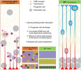
Combined removal of Crb1 and Crb2 from the developing optic vesicle evokes cellular and laminar disorganization by disrupting the apical cell-cell adhesion in developing retinal epithelium. As a result, at postnatal stages, affected mouse retinas show temporarily thickened, coarsely laminated retinas in addition to functional deficits, including a severely abnormal electroretinogram and decreased visual acuity. These features are reminiscent of Leber congenital amaurosis 8, which is caused in humans by subsets of Crb1 mutations. However, the cellular basis of the abnormalities in retinal progenitor cells (RPCs) that lead to retinal disorganization is largely unknown. In this study, we analyze specific features of RPCs in mutant retinas, including maintenance of the progenitor pool, cell cycle progression, cell cycle phase-dependent nuclear positioning, cell survival, and generation of mature retinal cell types. We find crucial defects in the mutant RPCs. Upon removal of CRB1 and CRB2, apical structures of the RPCs, determined by markers of cilia and centrosomes, are basally shifted. In addition, the positioning of the somata of the M-phase cells, normally localized at the apical surface of the retinal epithelium, is basally shifted in a nearly randomized pattern along the apico-basal axis. Consequently, we propose that positioning of RPCs is desynchronized from cell cycle phase and largely randomized during embryonic development at E17.5. Because the resultant postmitotic cells inevitably lose positional information, the outer and inner nuclear layers (ONL and INL) fail to form from ONBL during neonatal development and retinal cells become mixed locally and globally. Additional results of the lost tissue polarity in Crb1/Crb2 dKO retinas include atypical formation of heterotopic cell patches containing photoreceptor cells in the ganglion cell layer and acellular patches filled with neural processes. Collectively, these changes lead to a mouse model of LCA8-like pathology. LCA8-like pathology differs substantially from the well-characterized, broad range of degeneration phenotypes that arise during the differentiation of photoreceptor and Muller glial cells in retinitis pigmentosa 12, a closely related disease caused by mutated human Crb1.
Importantly, the present results suggest that Crb1/Crb2 serve indispensable functions in maintaining cell-cycle phase-dependent positioning of RPCs along the apico-basal axis, regulating cell cycle progression, and maintaining structural laminar integrity without significantly affecting the size of the RPC pools, generation of the subsets of the retinal cell types, or the distribution of cell cycle phases during RPC division. Taken together, these findings provide the crucial cellular basis of the thickening and severely disorganized lamination that are the unique features of the retinal abnormalities in LCA8 patients.

在 LCA8-like 小鼠模型中,视网膜祖细胞沿 apico-basal 轴的细胞周期相位依赖性定位和核迁移紊乱是全局性视网膜紊乱的基础。
通过破坏发育中视网膜上皮细胞顶端细胞-细胞间的粘附力,从发育中视囊中联合移除Crb1和Crb2会导致细胞和层状结构紊乱。因此,在出生后阶段,受影响的小鼠视网膜会出现暂时性增厚、粗层状结构,此外还会出现功能障碍,包括视网膜电图严重异常和视敏度下降。这些特征让人联想到因 Crb1 亚型突变而导致的 Leber 先天性无视力症 8。然而,视网膜祖细胞(RPC)的异常导致视网膜紊乱的细胞基础在很大程度上是未知的。在本研究中,我们分析了突变视网膜中 RPC 的具体特征,包括祖细胞池的维持、细胞周期的进展、细胞周期相位依赖性核定位、细胞存活以及成熟视网膜细胞类型的生成。我们发现突变体 RPC 存在关键缺陷。去除 CRB1 和 CRB2 后,由纤毛和中心体标记确定的 RPC 顶端结构发生基底移位。此外,通常位于视网膜上皮顶端表面的 M 期细胞的体节定位也沿顶端-基底轴以近乎随机的模式发生基底移动。因此,我们认为,在 E17.5 胚胎发育过程中,RPC 的定位与细胞周期阶段不同步,而且基本上是随机的。由于由此产生的有丝分裂后细胞不可避免地失去了位置信息,因此在新生儿发育过程中,核外层和核内层(ONL 和 INL)无法从 ONBL 形成,视网膜细胞在局部和整体上变得混杂。Crb1/Crb2 dKO 视网膜组织极性丧失的其他结果包括神经节细胞层中含有感光细胞的异位细胞斑块和充满神经过程的无细胞斑块的不典型形成。这些变化共同导致了一种 LCA8 类病理小鼠模型。LCA8样病理与视网膜色素变性12(一种由突变的人类Crb1引起的密切相关的疾病)中感光细胞和Muller神经胶质细胞分化过程中出现的特征明显、范围广泛的变性表型有很大不同。重要的是,本研究结果表明,Crb1/Crb2 在维持 RPC 沿 apico-basal 轴的细胞周期阶段性定位、调节细胞周期进程和维持结构板层完整性方面发挥着不可或缺的功能,而不会显著影响 RPC 池的大小、视网膜细胞类型亚群的生成或 RPC 分裂过程中细胞周期阶段的分布。综上所述,这些发现为 LCA8 患者视网膜增厚和层状结构严重紊乱提供了重要的细胞基础,而这正是 LCA8 患者视网膜异常的独特特征。
本文章由计算机程序翻译,如有差异,请以英文原文为准。
求助全文
约1分钟内获得全文
求助全文
来源期刊

Developmental biology
生物-发育生物学
CiteScore
5.30
自引率
3.70%
发文量
182
审稿时长
1.5 months
期刊介绍:
Developmental Biology (DB) publishes original research on mechanisms of development, differentiation, and growth in animals and plants at the molecular, cellular, genetic and evolutionary levels. Areas of particular emphasis include transcriptional control mechanisms, embryonic patterning, cell-cell interactions, growth factors and signal transduction, and regulatory hierarchies in developing plants and animals.
 求助内容:
求助内容: 应助结果提醒方式:


