KA-mediated excitotoxicity induces neuronal ferroptosis through activation of ferritinophagy

IF 4.8 1区 医学 Q1 NEUROSCIENCES
Yi-Yue Jiang, Wei-Long Wu, Jia-Ni Huang, Na Liu, Jing Wang, Xiao-Rui Wan, Zheng-Hong Qin, Yan Wang
{"title":"KA-mediated excitotoxicity induces neuronal ferroptosis through activation of ferritinophagy","authors":"Yi-Yue Jiang,&nbsp;Wei-Long Wu,&nbsp;Jia-Ni Huang,&nbsp;Na Liu,&nbsp;Jing Wang,&nbsp;Xiao-Rui Wan,&nbsp;Zheng-Hong Qin,&nbsp;Yan Wang","doi":"10.1111/cns.70054","DOIUrl":null,"url":null,"abstract":"<div>\n \n \n <section>\n \n <h3> Objectives</h3>\n \n <p>This study aims to elucidate the role of Fe<sup>2+</sup> overload in kainic acid (KA)-induced excitotoxicity, investigate the involvement of ferritinophagy selective cargo receptor NCOA4 in the pathogenesis of excitotoxicity.</p>\n </section>\n \n <section>\n \n <h3> Methods</h3>\n \n <p>Western blotting was used to detect the expression of FTH1, NCOA4, Lamp2, TfR, FPN, and DMT1 after KA stereotaxic injection into the unilateral striatum of mice. Colocalization of Fe<sup>2+</sup> with lysosomes in KA-treated primary cortical neurons was observed by using confocal microscopy. Desferrioxamine (DFO) was added to chelate free iron, a CCK8 kit was used to measure cell viability, and the Fe<sup>2+</sup> levels were detected by FerroOrange. BODIPY C11 was used to determine intracellular lipid reactive oxygen species (ROS) levels, and the mRNA levels of PTGS2, a biomarker of ferroptosis, were measured by fluorescent quantitative PCR. 3-Methyladenine (3-MA) was employed to inhibit KA-induced activation of autophagy, and changes in ferritinophagy-related protein expression and the indicated biomarkers of ferroptosis were detected. Endogenous NCOA4 was knocked down by lentivirus transfection, and cell viability and intracellular Fe<sup>2+</sup> levels were observed after KA treatment.</p>\n </section>\n \n <section>\n \n <h3> Results</h3>\n \n <p>Western blot results showed that the expression of NCOA4, DMT1, and Lamp2 was significantly upregulated, while FTH1 was downregulated, but there were no significant changes in TfR and FPN. The fluorescence results indicated that KA enhanced the colocalization of free Fe<sup>2+</sup> with lysosomes in neurons. DFO intervention could effectively rescue cell damage, reduce intracellular lipid peroxidation, and decrease the increased transcript levels of PTGS2 caused by KA. Pretreatment with 3-MA effectively reversed KA-induced ferritinophagy and ferroptosis. Endogenous interference with NCOA4 significantly improved cell viability and reduced intracellular free Fe<sup>2+</sup> levels in KA-treated cells.</p>\n </section>\n \n <section>\n \n <h3> Conclusion</h3>\n \n <p>KA-induced excitotoxicity activates ferritinophagy, and targeting ferritinophagy effectively inhibits downstream ferroptosis. Interference with NCOA4 effectively attenuates KA-induced neuronal damage. This study provides a potential therapeutic target for excitotoxicity related disease conditions.</p>\n </section>\n </div>","PeriodicalId":154,"journal":{"name":"CNS Neuroscience & Therapeutics","volume":"30 9","pages":""},"PeriodicalIF":4.8000,"publicationDate":"2024-09-22","publicationTypes":"Journal Article","fieldsOfStudy":null,"isOpenAccess":false,"openAccessPdf":"https://www.ncbi.nlm.nih.gov/pmc/articles/PMC11416743/pdf/","citationCount":"0","resultStr":null,"platform":"Semanticscholar","paperid":null,"PeriodicalName":"CNS Neuroscience & Therapeutics","FirstCategoryId":"3","ListUrlMain":"https://onlinelibrary.wiley.com/doi/10.1111/cns.70054","RegionNum":1,"RegionCategory":"医学","ArticlePicture":[],"TitleCN":null,"AbstractTextCN":null,"PMCID":null,"EPubDate":"","PubModel":"","JCR":"Q1","JCRName":"NEUROSCIENCES","Score":null,"Total":0}
引用次数: 0

Abstract

Objectives

This study aims to elucidate the role of Fe2+ overload in kainic acid (KA)-induced excitotoxicity, investigate the involvement of ferritinophagy selective cargo receptor NCOA4 in the pathogenesis of excitotoxicity.

Methods

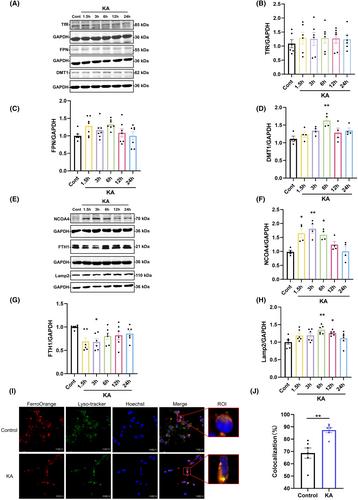
Western blotting was used to detect the expression of FTH1, NCOA4, Lamp2, TfR, FPN, and DMT1 after KA stereotaxic injection into the unilateral striatum of mice. Colocalization of Fe2+ with lysosomes in KA-treated primary cortical neurons was observed by using confocal microscopy. Desferrioxamine (DFO) was added to chelate free iron, a CCK8 kit was used to measure cell viability, and the Fe2+ levels were detected by FerroOrange. BODIPY C11 was used to determine intracellular lipid reactive oxygen species (ROS) levels, and the mRNA levels of PTGS2, a biomarker of ferroptosis, were measured by fluorescent quantitative PCR. 3-Methyladenine (3-MA) was employed to inhibit KA-induced activation of autophagy, and changes in ferritinophagy-related protein expression and the indicated biomarkers of ferroptosis were detected. Endogenous NCOA4 was knocked down by lentivirus transfection, and cell viability and intracellular Fe2+ levels were observed after KA treatment.

Results

Western blot results showed that the expression of NCOA4, DMT1, and Lamp2 was significantly upregulated, while FTH1 was downregulated, but there were no significant changes in TfR and FPN. The fluorescence results indicated that KA enhanced the colocalization of free Fe2+ with lysosomes in neurons. DFO intervention could effectively rescue cell damage, reduce intracellular lipid peroxidation, and decrease the increased transcript levels of PTGS2 caused by KA. Pretreatment with 3-MA effectively reversed KA-induced ferritinophagy and ferroptosis. Endogenous interference with NCOA4 significantly improved cell viability and reduced intracellular free Fe2+ levels in KA-treated cells.

Conclusion

KA-induced excitotoxicity activates ferritinophagy, and targeting ferritinophagy effectively inhibits downstream ferroptosis. Interference with NCOA4 effectively attenuates KA-induced neuronal damage. This study provides a potential therapeutic target for excitotoxicity related disease conditions.
Abstract Image

KA 介导的兴奋性毒性通过激活噬铁蛋白诱导神经元铁变态反应。
研究目的本研究旨在阐明Fe2+超载在凯尼酸(KA)诱导的兴奋性中毒中的作用,探讨噬铁选择性货物受体NCOA4参与兴奋性中毒的发病机制:方法:小鼠单侧纹状体注射 KA 后,采用 Western 印迹法检测 FTH1、NCOA4、Lamp2、TfR、FPN 和 DMT1 的表达。利用共聚焦显微镜观察了经 KA 处理的初级皮质神经元中 Fe2+ 与溶酶体的共定位。加入去铁胺(DFO)以螯合游离铁,使用 CCK8 试剂盒测量细胞活力,并用 FerroOrange 检测 Fe2+ 水平。用 BODIPY C11 测定细胞内脂质活性氧(ROS)水平,用荧光定量 PCR 检测铁变态反应生物标志物 PTGS2 的 mRNA 水平。采用 3-甲基腺嘌呤(3-MA)抑制 KA 诱导的自噬活化,并检测了嗜铁蛋白相关蛋白表达的变化和铁变态反应的指示生物标志物。通过慢病毒转染敲除内源性 NCOA4,观察 KA 处理后的细胞活力和细胞内 Fe2+ 水平:Western印迹结果显示,NCOA4、DMT1和Lamp2的表达明显上调,而FTH1的表达下调,但TfR和FPN的表达无明显变化。荧光结果表明,KA增强了游离Fe2+与神经元溶酶体的共定位。DFO干预能有效挽救细胞损伤,减少细胞内脂质过氧化,并降低KA引起的PTGS2转录水平的升高。3-MA预处理能有效逆转KA诱导的铁蛋白吞噬和铁突变。内源性干扰 NCOA4 能显著提高 KA 处理细胞的存活率并降低细胞内游离 Fe2+ 的水平:结论:KA诱导的兴奋性毒性激活了噬铁蛋白,针对噬铁蛋白能有效抑制下游的铁突变。干扰 NCOA4 能有效减轻 KA 诱导的神经元损伤。这项研究为兴奋毒性相关疾病提供了一个潜在的治疗靶点。
本文章由计算机程序翻译,如有差异,请以英文原文为准。
求助全文
约1分钟内获得全文 求助全文
来源期刊
CNS Neuroscience & Therapeutics
CNS Neuroscience & Therapeutics 医学-神经科学
CiteScore
7.30
自引率
12.70%
发文量
240
审稿时长
2 months
期刊介绍: CNS Neuroscience & Therapeutics provides a medium for rapid publication of original clinical, experimental, and translational research papers, timely reviews and reports of novel findings of therapeutic relevance to the central nervous system, as well as papers related to clinical pharmacology, drug development and novel methodologies for drug evaluation. The journal focuses on neurological and psychiatric diseases such as stroke, Parkinson’s disease, Alzheimer’s disease, depression, schizophrenia, epilepsy, and drug abuse.
×
引用
GB/T 7714-2015
复制
MLA
复制
APA
复制
导出至
BibTeX EndNote RefMan NoteFirst NoteExpress
×
提示
您的信息不完整,为了账户安全,请先补充。
现在去补充
×
提示
您因"违规操作"
具体请查看互助需知
我知道了
×
提示
确定
请完成安全验证×
copy
已复制链接
快去分享给好友吧!
我知道了
右上角分享
点击右上角分享
0
联系我们:info@booksci.cn Book学术提供免费学术资源搜索服务,方便国内外学者检索中英文文献。致力于提供最便捷和优质的服务体验。 Copyright © 2023 布克学术 All rights reserved.
京ICP备2023020795号-1
ghs 京公网安备 11010802042870号
Book学术文献互助
Book学术文献互助群
群 号:481959085
Book学术官方微信