Early clinico-radiological outcomes following neuroendoscopic cysto-cisternostomy for middle cranial fossa arachnoid cysts: a prospective cohort study with illustrative cases
Promise Tamunoipiriala Jaja, Yakimov Yuri, Albert Sufianov
{"title":"Early clinico-radiological outcomes following neuroendoscopic cysto-cisternostomy for middle cranial fossa arachnoid cysts: a prospective cohort study with illustrative cases","authors":"Promise Tamunoipiriala Jaja, Yakimov Yuri, Albert Sufianov","doi":"10.1007/s00381-024-06596-1","DOIUrl":null,"url":null,"abstract":"<h3 data-test=\"abstract-sub-heading\">Background</h3><p>The dysmorphogenetic arachnoid cysts’ pathomechanism is most favoured, and about 50% occur as middle cranial fossa cysts (MCFAC). Still being rare, management options are yet evolving. We described the clinico-radiological features, management and early outcomes of participants with MCFAC in our service.</p><h3 data-test=\"abstract-sub-heading\">Methods</h3><p>This prospective cohort study involved 29 pediatric participants recruited (from electronic health records, using ICD G93.0 D016080 for arachnoid cysts) between 01/01/2023 and 31/06/2023, following informed consent according to the ethical approval. All participants had neuro-imaging confirmed MCFAC. Baseline and follow-up data were retrieved and analyzed using summary (mean, standard deviation) and inferential (ANOVA, <i>t</i>-test) statistics.</p><h3 data-test=\"abstract-sub-heading\">Results</h3><p>They were averagely aged 6.2 ± 4.48 years and were mostly males (89.7%). 24.1% were asymptomatic. The commonest symptoms (<i>n</i> = 38) were headaches (23.7%), developmental delays (15.8%), eye complaints (15.8%) and cephalomegaly (7.9%). They were predominantly left-sided (89.7%). Galassi (G) 3 lesions were less (24.1%), with G2 and G1 lesions evenly sharing the rest. The average cyst volume was 58.4 ± 80.83cm<sup>3</sup>; there were significant differences (<i>F</i> = 4.682; <i>p</i> = 0.018) between the average volumes for G1 (14.4 ± 22.42cm<sup>3</sup>), G2 (61.7 ± 89.92cm<sup>3</sup>) and G3 (122.5 ± 94.37cm<sup>3</sup>) lesions. 44.8% of the participants had rigid-endoscopic cysto-cisternotomy (all between the ICA and oculomotor nerve into the interpeduncular cistern, using ventriculostomy forceps); including all G3, 50% of G2 and no G1 (had serial clinico-radiological observation) lesion. The average pre- (117.42cm<sup>3</sup>) and post-operative (53.48cm<sup>3</sup>) cyst volumes showed significant (<i>t</i> = − 2.797, <i>p</i> = 0.021) reductions.</p><h3 data-test=\"abstract-sub-heading\">Conclusion</h3><p>Middle cranial fossa arachnoid cysts occur predominantly amongst males, in middle childhood and left-sided. The treatment-related patient series are largely symptomatic, unlike the largely asymptomatic, screening-related series. Higher Galassi grade lesions presented with progressively, significantly larger cyst volumes and higher likelihoods of surgery. The average post-operative cyst volume at follow-up averagely showed almost 60% reduction from the pre-operative. All participants reported clinical remission.</p>","PeriodicalId":9970,"journal":{"name":"Child's Nervous System","volume":null,"pages":null},"PeriodicalIF":1.3000,"publicationDate":"2024-09-13","publicationTypes":"Journal Article","fieldsOfStudy":null,"isOpenAccess":false,"openAccessPdf":"","citationCount":"0","resultStr":null,"platform":"Semanticscholar","paperid":null,"PeriodicalName":"Child's Nervous System","FirstCategoryId":"3","ListUrlMain":"https://doi.org/10.1007/s00381-024-06596-1","RegionNum":4,"RegionCategory":"医学","ArticlePicture":[],"TitleCN":null,"AbstractTextCN":null,"PMCID":null,"EPubDate":"","PubModel":"","JCR":"Q4","JCRName":"CLINICAL NEUROLOGY","Score":null,"Total":0}
引用次数: 0
Abstract
Background
The dysmorphogenetic arachnoid cysts’ pathomechanism is most favoured, and about 50% occur as middle cranial fossa cysts (MCFAC). Still being rare, management options are yet evolving. We described the clinico-radiological features, management and early outcomes of participants with MCFAC in our service.
Methods
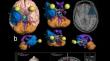
This prospective cohort study involved 29 pediatric participants recruited (from electronic health records, using ICD G93.0 D016080 for arachnoid cysts) between 01/01/2023 and 31/06/2023, following informed consent according to the ethical approval. All participants had neuro-imaging confirmed MCFAC. Baseline and follow-up data were retrieved and analyzed using summary (mean, standard deviation) and inferential (ANOVA, t-test) statistics.
Results
They were averagely aged 6.2 ± 4.48 years and were mostly males (89.7%). 24.1% were asymptomatic. The commonest symptoms (n = 38) were headaches (23.7%), developmental delays (15.8%), eye complaints (15.8%) and cephalomegaly (7.9%). They were predominantly left-sided (89.7%). Galassi (G) 3 lesions were less (24.1%), with G2 and G1 lesions evenly sharing the rest. The average cyst volume was 58.4 ± 80.83cm3; there were significant differences (F = 4.682; p = 0.018) between the average volumes for G1 (14.4 ± 22.42cm3), G2 (61.7 ± 89.92cm3) and G3 (122.5 ± 94.37cm3) lesions. 44.8% of the participants had rigid-endoscopic cysto-cisternotomy (all between the ICA and oculomotor nerve into the interpeduncular cistern, using ventriculostomy forceps); including all G3, 50% of G2 and no G1 (had serial clinico-radiological observation) lesion. The average pre- (117.42cm3) and post-operative (53.48cm3) cyst volumes showed significant (t = − 2.797, p = 0.021) reductions.
Conclusion
Middle cranial fossa arachnoid cysts occur predominantly amongst males, in middle childhood and left-sided. The treatment-related patient series are largely symptomatic, unlike the largely asymptomatic, screening-related series. Higher Galassi grade lesions presented with progressively, significantly larger cyst volumes and higher likelihoods of surgery. The average post-operative cyst volume at follow-up averagely showed almost 60% reduction from the pre-operative. All participants reported clinical remission.
期刊介绍:
The journal has been expanded to encompass all aspects of pediatric neurosciences concerning the developmental and acquired abnormalities of the nervous system and its coverings, functional disorders, epilepsy, spasticity, basic and clinical neuro-oncology, rehabilitation and trauma. Global pediatric neurosurgery is an additional field of interest that will be considered for publication in the journal.
 求助内容:
求助内容: 应助结果提醒方式:


