Preparation and Biological Evaluation of Porous Tantalum Scaffolds Coated with Hydroxyapatite
IF 4.6
Q2 MATERIALS SCIENCE, BIOMATERIALS
引用次数: 0
Abstract
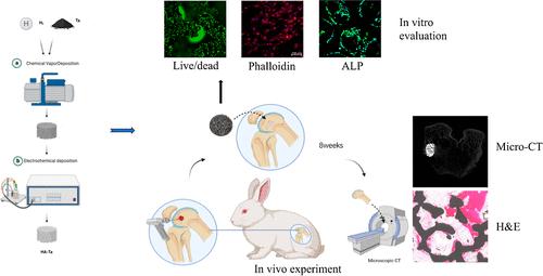
Orthopedic implants, such as porous scaffolds, are an effective way to repair bone defects. However, the lack of osseointegration and osteoinduction limits the achievement of an ideal therapeutic effect. This study aimed to prepare hydroxyapatite (HA) coatings for the surface of porous tantalum (Ta) scaffolds and to assess the effectively improved biological activities of the coated scaffolds. The porous Ta scaffolds were prepared by chemical vapor deposition, and then the porous Ta scaffolds were coated with HA via electrochemical deposition. The elements and phase compositions of the coatings were analyzed by energy-dispersive X-ray spectroscopy, X-ray diffraction, and X-ray photoelectron spectroscopy. The results showed that the coating covered the whole surfaces of porous Ta scaffolds with a uniform and compact distribution and did not exert any obvious effect on the porous structure. The biological activity of porous Ta scaffolds after surface modification increased and the water contact angle decreased, indicating that hydrophilicity was significantly improved. Cell live/dead staining, cytoskeletal fluorescence staining, and alkaline phosphatase immunofluorescence staining showed that the coating exhibited no cytotoxicity and notably improved cell proliferation, spreading, and osteogenic differentiation. In addition, in vivo experiments in animals have demonstrated that HA-coated porous Ta scaffolds contribute to bone formation. In conclusion, the HA coating notably improves the biological activities of the porous Ta scaffolds, achieving the goal of the present study. The HA coating presents great potential for the modification of porous Ta implants to improve their osteogenesis and osseointegration.

涂有羟基磷灰石的多孔钽支架的制备与生物学评估
多孔支架等骨科植入物是修复骨缺损的有效方法。然而,由于缺乏骨结合和骨诱导,限制了理想治疗效果的实现。本研究旨在为多孔钽(Ta)支架表面制备羟基磷灰石(HA)涂层,并评估涂层支架有效改善的生物活性。实验采用化学气相沉积法制备多孔钽支架,然后通过电化学沉积法在多孔钽支架表面涂覆HA。通过能量色散 X 射线光谱、X 射线衍射和 X 射线光电子能谱分析了涂层的元素和相组成。结果表明,涂层覆盖了多孔钽支架的整个表面,且分布均匀、紧密,对多孔结构无明显影响。表面改性后多孔钽支架的生物活性提高,水接触角减小,表明亲水性明显改善。细胞活/死染色、细胞骨架荧光染色和碱性磷酸酶免疫荧光染色表明,涂层无细胞毒性,并明显改善了细胞增殖、扩散和成骨分化。此外,动物体内实验表明,HA 涂层多孔 Ta 支架有助于骨形成。总之,HA 涂层显著提高了多孔 Ta 支架的生物活性,实现了本研究的目标。HA 涂层在改良多孔 Ta 植入物以改善其骨生成和骨整合方面具有巨大潜力。
本文章由计算机程序翻译,如有差异,请以英文原文为准。
求助全文
约1分钟内获得全文
求助全文
来源期刊

ACS Applied Bio Materials
Chemistry-Chemistry (all)
CiteScore
9.40
自引率
2.10%
发文量
464
期刊介绍:
ACS Applied Bio Materials is an interdisciplinary journal publishing original research covering all aspects of biomaterials and biointerfaces including and beyond the traditional biosensing, biomedical and therapeutic applications.
The journal is devoted to reports of new and original experimental and theoretical research of an applied nature that integrates knowledge in the areas of materials, engineering, physics, bioscience, and chemistry into important bio applications. The journal is specifically interested in work that addresses the relationship between structure and function and assesses the stability and degradation of materials under relevant environmental and biological conditions.
 求助内容:
求助内容: 应助结果提醒方式:


