Neural responses to camouflage targets with different exposure signs based on EEG

IF 4.6 Q2 MATERIALS SCIENCE, BIOMATERIALS
Zhou Yu , Li Xue , Weidong Xu , Jun Liu , Qi Jia , Yawen Liu , Lu Zhou , Jianghua Hu , Hao Li , Jidong Wu
{"title":"Neural responses to camouflage targets with different exposure signs based on EEG","authors":"Zhou Yu ,&nbsp;Li Xue ,&nbsp;Weidong Xu ,&nbsp;Jun Liu ,&nbsp;Qi Jia ,&nbsp;Yawen Liu ,&nbsp;Lu Zhou ,&nbsp;Jianghua Hu ,&nbsp;Hao Li ,&nbsp;Jidong Wu","doi":"10.1016/j.neuropsychologia.2024.109002","DOIUrl":null,"url":null,"abstract":"<div><p>This study investigates the relationship between various target exposure signs and brain activation patterns by analyzing the EEG signals of 35 subjects observing four types of targets: well-camouflaged, with large color differences, with shadows, and of large size. Through ERP analysis and source localization, we have established that different exposure signs elicit distinct brain activation patterns. The ERP analysis revealed a strong correlation between the latency of the P300 component and the visibility of the exposure signs. Furthermore, our source localization findings indicate that exposure signs alter the current density distribution within the cortex, with shadows causing significantly higher activation in the frontal lobe compared to other conditions. The study also uncovered a pronounced right-brain laterality in subjects during target identification. By employing an LSTM neural network, we successfully differentiated EEG signals triggered by various exposure signs, achieving a classification accuracy of up to 96.4%. These results not only suggest that analyzing the P300 latency and cortical current distribution can differentiate the degree of visibility of target exposure signs, but also demonstrate the potential of using EEG characteristics to identify key exposure signs in camouflaged targets. This provides crucial insights for developing auxiliary camouflage strategies.</p></div>","PeriodicalId":2,"journal":{"name":"ACS Applied Bio Materials","volume":null,"pages":null},"PeriodicalIF":4.6000,"publicationDate":"2024-09-16","publicationTypes":"Journal Article","fieldsOfStudy":null,"isOpenAccess":false,"openAccessPdf":"","citationCount":"0","resultStr":null,"platform":"Semanticscholar","paperid":null,"PeriodicalName":"ACS Applied Bio Materials","FirstCategoryId":"102","ListUrlMain":"https://www.sciencedirect.com/science/article/pii/S0028393224002173","RegionNum":0,"RegionCategory":null,"ArticlePicture":[],"TitleCN":null,"AbstractTextCN":null,"PMCID":null,"EPubDate":"","PubModel":"","JCR":"Q2","JCRName":"MATERIALS SCIENCE, BIOMATERIALS","Score":null,"Total":0}
引用次数: 0

Abstract

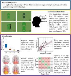
This study investigates the relationship between various target exposure signs and brain activation patterns by analyzing the EEG signals of 35 subjects observing four types of targets: well-camouflaged, with large color differences, with shadows, and of large size. Through ERP analysis and source localization, we have established that different exposure signs elicit distinct brain activation patterns. The ERP analysis revealed a strong correlation between the latency of the P300 component and the visibility of the exposure signs. Furthermore, our source localization findings indicate that exposure signs alter the current density distribution within the cortex, with shadows causing significantly higher activation in the frontal lobe compared to other conditions. The study also uncovered a pronounced right-brain laterality in subjects during target identification. By employing an LSTM neural network, we successfully differentiated EEG signals triggered by various exposure signs, achieving a classification accuracy of up to 96.4%. These results not only suggest that analyzing the P300 latency and cortical current distribution can differentiate the degree of visibility of target exposure signs, but also demonstrate the potential of using EEG characteristics to identify key exposure signs in camouflaged targets. This provides crucial insights for developing auxiliary camouflage strategies.
Abstract Image

基于脑电图的对不同曝光标志的伪装目标的神经反应
本研究通过分析 35 名受试者在观察伪装良好、色差较大、有阴影和体积较大四种类型目标时的脑电信号,研究了各种目标暴露标志与大脑激活模式之间的关系。通过ERP分析和信号源定位,我们确定了不同的暴露标志会引起不同的大脑激活模式。ERP分析显示,P300分量的潜伏期与曝光标志的可见度之间存在很强的相关性。此外,我们的源定位研究结果表明,曝光标志改变了大脑皮层内的电流密度分布,与其他条件相比,阴影导致额叶的激活明显更高。研究还发现,受试者在目标识别过程中存在明显的右脑侧向性。通过使用 LSTM 神经网络,我们成功地区分了由各种曝光迹象引发的脑电信号,分类准确率高达 96.4%。这些结果不仅表明,分析 P300 潜伏期和皮层电流分布可以区分目标暴露标志的可见度,还证明了利用脑电图特征识别伪装目标中关键暴露标志的潜力。这为开发辅助伪装策略提供了重要的启示。
本文章由计算机程序翻译,如有差异,请以英文原文为准。
求助全文
约1分钟内获得全文 求助全文
来源期刊
ACS Applied Bio Materials
ACS Applied Bio Materials Chemistry-Chemistry (all)
CiteScore
9.40
自引率
2.10%
发文量
464
×
引用
GB/T 7714-2015
复制
MLA
复制
APA
复制
导出至
BibTeX EndNote RefMan NoteFirst NoteExpress
×
提示
您的信息不完整,为了账户安全,请先补充。
现在去补充
×
提示
您因"违规操作"
具体请查看互助需知
我知道了
×
提示
确定
请完成安全验证×
copy
已复制链接
快去分享给好友吧!
我知道了
右上角分享
点击右上角分享
0
联系我们:info@booksci.cn Book学术提供免费学术资源搜索服务,方便国内外学者检索中英文文献。致力于提供最便捷和优质的服务体验。 Copyright © 2023 布克学术 All rights reserved.
京ICP备2023020795号-1
ghs 京公网安备 11010802042870号
Book学术文献互助
Book学术文献互助群
群 号:481959085
Book学术官方微信