Courtney P. Korff, Sophie Nelissen, Amy B. Todd-Donato, Andrew D. Miller, Emma Davies
{"title":"Malignant transformation of an intraparenchymal hemangioma in the cervical spinal cord of a German shepherd dog","authors":"Courtney P. Korff, Sophie Nelissen, Amy B. Todd-Donato, Andrew D. Miller, Emma Davies","doi":"10.1111/jvim.17190","DOIUrl":null,"url":null,"abstract":"<p>An 8-year-old female spayed German shepherd dog was presented for evaluation of a 1-week history of right thoracic limb monoparesis. Magnetic resonance imaging (MRI) identified an intraparenchymal, T2 hypointense and T1 isointense, strongly heterogeneously contrast-enhancing mass with moderate internal susceptibility artifact on T2* images at the level of the cranial extent of the C5 vertebral body. Euthanasia was elected after a rapid neurologic decline in the 24 hours after MRI. Necropsy and histopathology identified an intraparenchymal hemangiosarcoma arising from a hemangioma in the cervical spinal cord, with no evidence of neoplastic disease in any other examined organs. The spectrum of vasoproliferative disorders in the central nervous system in veterinary species has been codified recently, but hemangiosarcoma is considered metastatic to the central nervous system. Herein we describe the clinical, imaging, and histologic findings in a dog with a novel primary location of hemangiosarcoma in the cervical spinal cord.</p>","PeriodicalId":49958,"journal":{"name":"Journal of Veterinary Internal Medicine","volume":"38 5","pages":"2681-2685"},"PeriodicalIF":2.1000,"publicationDate":"2024-09-11","publicationTypes":"Journal Article","fieldsOfStudy":null,"isOpenAccess":false,"openAccessPdf":"https://onlinelibrary.wiley.com/doi/epdf/10.1111/jvim.17190","citationCount":"0","resultStr":null,"platform":"Semanticscholar","paperid":null,"PeriodicalName":"Journal of Veterinary Internal Medicine","FirstCategoryId":"97","ListUrlMain":"https://onlinelibrary.wiley.com/doi/10.1111/jvim.17190","RegionNum":2,"RegionCategory":"农林科学","ArticlePicture":[],"TitleCN":null,"AbstractTextCN":null,"PMCID":null,"EPubDate":"","PubModel":"","JCR":"Q1","JCRName":"VETERINARY SCIENCES","Score":null,"Total":0}
引用次数: 0
Abstract
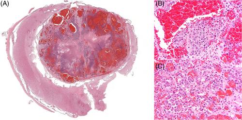
An 8-year-old female spayed German shepherd dog was presented for evaluation of a 1-week history of right thoracic limb monoparesis. Magnetic resonance imaging (MRI) identified an intraparenchymal, T2 hypointense and T1 isointense, strongly heterogeneously contrast-enhancing mass with moderate internal susceptibility artifact on T2* images at the level of the cranial extent of the C5 vertebral body. Euthanasia was elected after a rapid neurologic decline in the 24 hours after MRI. Necropsy and histopathology identified an intraparenchymal hemangiosarcoma arising from a hemangioma in the cervical spinal cord, with no evidence of neoplastic disease in any other examined organs. The spectrum of vasoproliferative disorders in the central nervous system in veterinary species has been codified recently, but hemangiosarcoma is considered metastatic to the central nervous system. Herein we describe the clinical, imaging, and histologic findings in a dog with a novel primary location of hemangiosarcoma in the cervical spinal cord.
期刊介绍:
The mission of the Journal of Veterinary Internal Medicine is to advance veterinary medical knowledge and improve the lives of animals by publication of authoritative scientific articles of animal diseases.
 求助内容:
求助内容: 应助结果提醒方式:


