Frontiers in artificial intelligence-directed light-sheet microscopy for uncovering biological phenomena and multiorgan imaging
IF 8.5
4区 医学
Q1 MATERIALS SCIENCE, BIOMATERIALS
引用次数: 0
Abstract
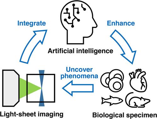
Light-sheet fluorescence microscopy (LSFM) introduces fast scanning of biological phenomena with deep photon penetration and minimal phototoxicity. This advancement represents a significant shift in 3-D imaging of large-scale biological tissues and 4-D (space + time) imaging of small live animals. The large data associated with LSFM require efficient imaging acquisition and analysis with the use of artificial intelligence (AI)/machine learning (ML) algorithms. To this end, AI/ML-directed LSFM is an emerging area for multiorgan imaging and tumor diagnostics. This review will present the development of LSFM and highlight various LSFM configurations and designs for multiscale imaging. Optical clearance techniques will be compared for effective reduction in light scattering and optimal deep-tissue imaging. This review will further depict a diverse range of research and translational applications, from small live organisms to multiorgan imaging to tumor diagnosis. In addition, this review will address AI/ML-directed imaging reconstruction, including the application of convolutional neural networks (CNNs) and generative adversarial networks (GANs). In summary, the advancements of LSFM have enabled effective and efficient post-imaging reconstruction and data analyses, underscoring LSFM's contribution to advancing fundamental and translational research.
用于揭示生物现象和多器官成像的人工智能定向光片显微镜的前沿领域
光片荧光显微镜(LSFM)可对生物现象进行快速扫描,光子穿透力强,光毒性极低。这一进步标志着大规模生物组织三维成像和小型活体动物四维(空间+时间)成像的重大转变。与 LSFM 相关的大数据需要利用人工智能/机器学习算法进行高效的成像采集和分析。为此,人工智能/ML 引导的 LSFM 是多器官成像和肿瘤诊断的一个新兴领域。本综述将介绍 LSFM 的发展,并重点介绍用于多尺度成像的各种 LSFM 配置和设计。将对光学清除技术进行比较,以有效减少光散射和优化深部组织成像。本综述将进一步描述从小型活体到多器官成像再到肿瘤诊断的各种研究和转化应用。此外,本综述还将讨论人工智能/ML 引导的成像重建,包括卷积神经网络(CNN)和生成对抗网络(GAN)的应用。总之,LSFM 的进步实现了有效和高效的成像后重建和数据分析,凸显了 LSFM 在推动基础研究和转化研究方面的贡献。
本文章由计算机程序翻译,如有差异,请以英文原文为准。
求助全文
约1分钟内获得全文
求助全文
来源期刊
VIEW
Multiple-
CiteScore
12.60
自引率
2.30%
发文量
0
审稿时长
10 weeks
期刊介绍:
View publishes scientific articles studying novel crucial contributions in the areas of Biomaterials and General Chemistry. View features original academic papers which go through peer review by experts in the given subject area.View encourages submissions from the research community where the priority will be on the originality and the practical impact of the reported research.
 求助内容:
求助内容: 应助结果提醒方式:


