Periaqueductal gray connectivity in spinal cord injury-induced neuropathic pain
Abstract
Background and Purpose
Neuropathic pain (NP) is a debilitating condition following spinal cord injury (SCI). The role of periaqueductal gray (PAG) in NP development following SCI remains underexplored. Using resting-state functional MRI (rsfMRI), our study aimed to demonstrate the alterations in functional connectivity (FC) of PAG in NP following SCI.
Methods
Ten SCI patients (SCI + NP, n = 7, and SCI − NP, n = 3), alongside 10 healthy controls (HCs), were enrolled. rsfMRI was conducted followed by seed-to-voxel analysis using PAG as the seed region and then group-based analysis comprising three groups (SCI + NP, SCI − NP, and HC). Age and gender were considered as confounding variables.
Results
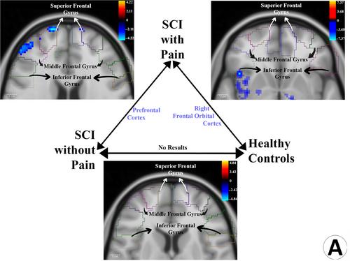
Compared to HCs, SCI + NP demonstrated decreased FC between PAG and right insula, right frontal orbital cortex, right pallidum, dorsal raphe nucleus (DRN), red nuclei (RN), substantia nigra (SN), and ventral posterolateral (VPL) thalamic nuclei. Compared to SCI − NP, SCI + NP demonstrated increased FC between PAG and posterior cingulate cortex (PCC), hippocampus, cerebellar vermis lobules IV and V, and thalamic structures (posterior and lateral pulvinar, the mediodorsal nuclei, and the ventral lateral nuclei). Additionally, decreased FC between the PAG and VPL, geniculate bodies, intralaminar nuclei of thalamus, DRN, RN, SN, and prefrontal cortex was observed in this comparison.
Conclusions
Altered FC between PAG and right anterior insula, VPL, DRN, RN, SN, cerebellar vermis lobules IV and V, frontal cortex, and PCC was associated with NP sequelae of SCI. Additionally, SCI was independently associated with decreased FC between PAG and right posterior insula, cerebellar lobules IV and V, and cerebellar vermis lobules III, IV, and V.

 求助内容:
求助内容: 应助结果提醒方式:


