{"title":"In situ interface reaction-enabled electrochemiluminescence imaging for single-cell formaldehyde release analysis†","authors":"Juanhua Zhou and Yang Liu","doi":"10.1039/D4SD00177J","DOIUrl":null,"url":null,"abstract":"<p >Monitoring metabolites <em>in situ</em> at the single-cell scale is important for revealing cellular heterogeneity and dynamic changes of cell status, which provides new possibilities for disease research. Benefiting from the advantages of both electrochemical and optical methods, electrochemiluminescence (ECL) has great potential in this field. However, developing real-time <em>in situ</em> imaging methods is full of challenges. In this study, an ECL imaging method for formaldehyde (FA), a kind of cellular metabolite, was developed based on the <em>in situ</em> generation of co-reactants at the electrode interface and was successfully applied to the monitoring of single-cell FA release. Amino groups can undergo a rapid nucleophilic addition reaction with FA to form amino alcohol intermediates, which can be used as co-reactants for tris(2,2′-bipyridyl)ruthenium(<small>II</small>) [Ru(bpy)<small><sub>3</sub></small><small><sup>2+</sup></small>] to significantly enhance the strength of ECL. Poly(amidoamine) (PAMAM), with a large number of amino groups, and reduced graphene oxide (rGO), with excellent electrical conductivity and electrocatalytic properties, were introduced as the modification layer on the electrode surface to realize the “turn on” detection of FA. This sensing method also eliminated the use of the classic toxic co-reactant tripropylamine (TPrA) and was further applied to <em>in situ</em> imaging of single-cell FA release. It successfully obtained ECL images at different time points after the stimulation of HeLa cells with thapsigargin (TG), revealing the change pattern in drug efficacy over time. This work proposes a new ECL imaging approach for real-time <em>in situ</em> monitoring of FA release from single cells, further broadening the application of ECL imaging in single-cell analysis.</p>","PeriodicalId":74786,"journal":{"name":"Sensors & diagnostics","volume":" 9","pages":" 1571-1578"},"PeriodicalIF":4.1000,"publicationDate":"2024-08-20","publicationTypes":"Journal Article","fieldsOfStudy":null,"isOpenAccess":false,"openAccessPdf":"https://pubs.rsc.org/en/content/articlepdf/2024/sd/d4sd00177j?page=search","citationCount":"0","resultStr":null,"platform":"Semanticscholar","paperid":null,"PeriodicalName":"Sensors & diagnostics","FirstCategoryId":"1085","ListUrlMain":"https://pubs.rsc.org/en/content/articlelanding/2024/sd/d4sd00177j","RegionNum":0,"RegionCategory":null,"ArticlePicture":[],"TitleCN":null,"AbstractTextCN":null,"PMCID":null,"EPubDate":"","PubModel":"","JCR":"Q2","JCRName":"CHEMISTRY, ANALYTICAL","Score":null,"Total":0}
引用次数: 0
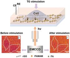
Abstract
Monitoring metabolites in situ at the single-cell scale is important for revealing cellular heterogeneity and dynamic changes of cell status, which provides new possibilities for disease research. Benefiting from the advantages of both electrochemical and optical methods, electrochemiluminescence (ECL) has great potential in this field. However, developing real-time in situ imaging methods is full of challenges. In this study, an ECL imaging method for formaldehyde (FA), a kind of cellular metabolite, was developed based on the in situ generation of co-reactants at the electrode interface and was successfully applied to the monitoring of single-cell FA release. Amino groups can undergo a rapid nucleophilic addition reaction with FA to form amino alcohol intermediates, which can be used as co-reactants for tris(2,2′-bipyridyl)ruthenium(II) [Ru(bpy)32+] to significantly enhance the strength of ECL. Poly(amidoamine) (PAMAM), with a large number of amino groups, and reduced graphene oxide (rGO), with excellent electrical conductivity and electrocatalytic properties, were introduced as the modification layer on the electrode surface to realize the “turn on” detection of FA. This sensing method also eliminated the use of the classic toxic co-reactant tripropylamine (TPrA) and was further applied to in situ imaging of single-cell FA release. It successfully obtained ECL images at different time points after the stimulation of HeLa cells with thapsigargin (TG), revealing the change pattern in drug efficacy over time. This work proposes a new ECL imaging approach for real-time in situ monitoring of FA release from single cells, further broadening the application of ECL imaging in single-cell analysis.
在单细胞尺度上原位监测代谢物对于揭示细胞异质性和细胞状态的动态变化非常重要,这为疾病研究提供了新的可能性。得益于电化学和光学方法的优势,电化学发光(ECL)在这一领域具有巨大潜力。然而,开发实时原位成像方法充满挑战。本研究基于电极界面原位生成共反应物的原理,开发了一种针对细胞代谢物甲醛(FA)的 ECL 成像方法,并成功应用于单细胞 FA 释放的监测。氨基可与 FA 发生快速亲核加成反应,形成氨基醇中间体,这些中间体可用作三(2,2′-联吡啶)钌(II) [Ru(bpy)32+] 的共反应物,从而显著增强 ECL 的强度。电极表面引入了具有大量氨基的聚氨基胺(PAMAM)和具有优异导电性和电催化性能的还原氧化石墨烯(rGO)作为修饰层,实现了 FA 的 "开启 "检测。这种传感方法还省去了传统的有毒共反应物三丙胺(TPrA),并进一步应用于单细胞 FA 释放的原位成像。该方法成功地获得了用硫代甘氨酸(TG)刺激 HeLa 细胞后不同时间点的 ECL 图像,揭示了药效随时间的变化规律。这项工作提出了一种新的 ECL 成像方法,用于实时原位监测单细胞中 FA 的释放,进一步拓宽了 ECL 成像在单细胞分析中的应用。

 求助内容:
求助内容: 应助结果提醒方式:


