Impact of different parametric Patlak imaging approaches and comparison with a 2-tissue compartment pharmacokinetic model with a long axial field-of-view (LAFOV) PET/CT in oncological patients
IF 8.6 1区 医学Q1 RADIOLOGY, NUCLEAR MEDICINE & MEDICAL IMAGING
{"title":"Impact of different parametric Patlak imaging approaches and comparison with a 2-tissue compartment pharmacokinetic model with a long axial field-of-view (LAFOV) PET/CT in oncological patients","authors":"Leyun Pan, Christos Sachpekidis, Jessica Hassel, Petros Christopoulos, Antonia Dimitrakopoulou-Strauss","doi":"10.1007/s00259-024-06879-4","DOIUrl":null,"url":null,"abstract":"<h3 data-test=\"abstract-sub-heading\">Aim</h3><p>The recently introduced Long-Axial-Field-of-View (LAFOV) PET-CT scanners allow for the first-time whole-body dynamic- and parametric imaging. Primary aim of this study was the comparison of direct and indirect Patlak imaging as well as the comparison of different time frames for Patlak calculation with the LAFOV PET-CT in oncological patients. Secondary aims of the study were lesion detectability and comparison of Patlak analysis with a two-tissue-compartment model (2TCM).</p><h3 data-test=\"abstract-sub-heading\">Methodology</h3><p>50 oncological patients with 346 tumor lesions were enrolled in the study. All patients underwent [<sup>18</sup>F]FDG PET/CT (skull to upper thigh). Here, the Image-Derived-Input-Function) (IDIF) from the descending aorta was used as the exclusive input function. Four sets of images have been reviewed visually and evaluated quantitatively using the target-to-background (TBR) and contrast-to-noise ratio (CNR): short-time (30 min)-direct (STD) Patlak K<sub>i</sub>, short-time (30 min)-indirect (STI) Patlak K<sub>i</sub>, long-time (59.25 min)-indirect (LTI) Patlak K<sub>i</sub>, and 50–60 min SUV (sumSUV). VOI-based 2TCM was used for the evaluation of tumor lesions and normal tissues and compared with the results of Patlak model.</p><h3 data-test=\"abstract-sub-heading\">Results</h3><p>No significant differences were observed between the four approaches regarding the number of tumor lesions. However, we found three discordant results: a true positive liver lesion in all Patlak K<sub>i</sub> images, a false positive liver lesion delineated only in LTI K<sub>i</sub> which was a hemangioma according to MRI and a true negative example in a patient with an atelectasis next to a lung tumor. STD, STI and LTI K<sub>i</sub> images had superior TBR in comparison with sumSUV images (2.9-, 3.3- and 4.3-fold higher respectively). TBR of LTI K<sub>i</sub> were significantly higher than STD K<sub>i</sub>. VOI-based k<sub>3</sub> showed a 21-fold higher TBR than sumSUV. Parameters of different models vary in their differential capability between tumor lesions and normal tissue like Patlak K<sub>i</sub> which was better in normal lung and 2TCM k<sub>3</sub> which was better in normal liver. 2TCM K<sub>i</sub> revealed the highest correlation (<i>r</i> = 0.95) with the LTI Patlak K<sub>i</sub> in tumor lesions group and demonstrated the highest correlation with the STD Patlak K<sub>i</sub> in all tissues group and normal tissues group (<i>r</i> = 0.93 and <i>r</i> = 0.74 respectively).</p><h3 data-test=\"abstract-sub-heading\">Conclusions</h3><p>Dynamic [<sup>18</sup>F]-FDG with the new LAFOV PET/CT scanner produces Patlak K<sub>i</sub> images with better lesion contrast than SUV images, but does not increase the lesion detection rate. The time window used for Patlak imaging plays a more important role than the direct or indirect method. A combination of different models, like Patlak and 2TCM may be helpful in parametric imaging to obtain the best TBR in the whole body in future.</p>","PeriodicalId":11909,"journal":{"name":"European Journal of Nuclear Medicine and Molecular Imaging","volume":null,"pages":null},"PeriodicalIF":8.6000,"publicationDate":"2024-09-11","publicationTypes":"Journal Article","fieldsOfStudy":null,"isOpenAccess":false,"openAccessPdf":"","citationCount":"0","resultStr":null,"platform":"Semanticscholar","paperid":null,"PeriodicalName":"European Journal of Nuclear Medicine and Molecular Imaging","FirstCategoryId":"3","ListUrlMain":"https://doi.org/10.1007/s00259-024-06879-4","RegionNum":1,"RegionCategory":"医学","ArticlePicture":[],"TitleCN":null,"AbstractTextCN":null,"PMCID":null,"EPubDate":"","PubModel":"","JCR":"Q1","JCRName":"RADIOLOGY, NUCLEAR MEDICINE & MEDICAL IMAGING","Score":null,"Total":0}
引用次数: 0
Abstract
Aim
The recently introduced Long-Axial-Field-of-View (LAFOV) PET-CT scanners allow for the first-time whole-body dynamic- and parametric imaging. Primary aim of this study was the comparison of direct and indirect Patlak imaging as well as the comparison of different time frames for Patlak calculation with the LAFOV PET-CT in oncological patients. Secondary aims of the study were lesion detectability and comparison of Patlak analysis with a two-tissue-compartment model (2TCM).
Methodology
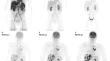
50 oncological patients with 346 tumor lesions were enrolled in the study. All patients underwent [18F]FDG PET/CT (skull to upper thigh). Here, the Image-Derived-Input-Function) (IDIF) from the descending aorta was used as the exclusive input function. Four sets of images have been reviewed visually and evaluated quantitatively using the target-to-background (TBR) and contrast-to-noise ratio (CNR): short-time (30 min)-direct (STD) Patlak Ki, short-time (30 min)-indirect (STI) Patlak Ki, long-time (59.25 min)-indirect (LTI) Patlak Ki, and 50–60 min SUV (sumSUV). VOI-based 2TCM was used for the evaluation of tumor lesions and normal tissues and compared with the results of Patlak model.
Results
No significant differences were observed between the four approaches regarding the number of tumor lesions. However, we found three discordant results: a true positive liver lesion in all Patlak Ki images, a false positive liver lesion delineated only in LTI Ki which was a hemangioma according to MRI and a true negative example in a patient with an atelectasis next to a lung tumor. STD, STI and LTI Ki images had superior TBR in comparison with sumSUV images (2.9-, 3.3- and 4.3-fold higher respectively). TBR of LTI Ki were significantly higher than STD Ki. VOI-based k3 showed a 21-fold higher TBR than sumSUV. Parameters of different models vary in their differential capability between tumor lesions and normal tissue like Patlak Ki which was better in normal lung and 2TCM k3 which was better in normal liver. 2TCM Ki revealed the highest correlation (r = 0.95) with the LTI Patlak Ki in tumor lesions group and demonstrated the highest correlation with the STD Patlak Ki in all tissues group and normal tissues group (r = 0.93 and r = 0.74 respectively).
Conclusions
Dynamic [18F]-FDG with the new LAFOV PET/CT scanner produces Patlak Ki images with better lesion contrast than SUV images, but does not increase the lesion detection rate. The time window used for Patlak imaging plays a more important role than the direct or indirect method. A combination of different models, like Patlak and 2TCM may be helpful in parametric imaging to obtain the best TBR in the whole body in future.
期刊介绍:
The European Journal of Nuclear Medicine and Molecular Imaging serves as a platform for the exchange of clinical and scientific information within nuclear medicine and related professions. It welcomes international submissions from professionals involved in the functional, metabolic, and molecular investigation of diseases. The journal's coverage spans physics, dosimetry, radiation biology, radiochemistry, and pharmacy, providing high-quality peer review by experts in the field. Known for highly cited and downloaded articles, it ensures global visibility for research work and is part of the EJNMMI journal family.

 求助内容:
求助内容: 应助结果提醒方式:


