Dietary fiber promotes antigen presentation on intestinal epithelial cells and development of small intestinal CD4+CD8αα+ intraepithelial T cells.

IF 7.9 2区 医学 Q1 IMMUNOLOGY
Naomi Rodriguez-Marino, Charlotte J Royer, Dormarie E Rivera-Rodriguez, Emma Seto, Isabelle Gracien, Rheinallt M Jones, Christopher D Scharer, Adam D Gracz, Luisa Cervantes-Barragan
{"title":"Dietary fiber promotes antigen presentation on intestinal epithelial cells and development of small intestinal CD4<sup>+</sup>CD8αα<sup>+</sup> intraepithelial T cells.","authors":"Naomi Rodriguez-Marino, Charlotte J Royer, Dormarie E Rivera-Rodriguez, Emma Seto, Isabelle Gracien, Rheinallt M Jones, Christopher D Scharer, Adam D Gracz, Luisa Cervantes-Barragan","doi":"10.1016/j.mucimm.2024.08.010","DOIUrl":null,"url":null,"abstract":"<p><p>The impact of dietary fiber on intestinal T cell development is poorly understood. Here we show that a low fiber diet reduces MHC-II antigen presentation by small intestinal epithelial cells (IECs) and consequently impairs development of CD4<sup>+</sup>CD8αα<sup>+</sup> intraepithelial lymphocytes (DP IELs) through changes to the microbiota. Dietary fiber supports colonization by Segmented Filamentous Bacteria (SFB), which induces the secretion of IFNγ by type 1 innate lymphoid cells (ILC1s) that lead to MHC-II upregulation on IECs. IEC MHC-II expression caused either by SFB colonization or exogenous IFNγ administration induced differentiation of DP IELs. Finally, we show that a low fiber diet promotes overgrowth of Bifidobacterium pseudolongum, and that oral administration of B. pseudolongum reduces SFB abundance in the small intestine. Collectively we highlight the importance of dietary fiber in maintaining the balance among microbiota members that allow IEC MHC-II antigen presentation and define a mechanism of microbiota-ILC-IEC interactions participating in the development of intestinal intraepithelial T cells.</p>","PeriodicalId":18877,"journal":{"name":"Mucosal Immunology","volume":" ","pages":""},"PeriodicalIF":7.9000,"publicationDate":"2024-09-05","publicationTypes":"Journal Article","fieldsOfStudy":null,"isOpenAccess":false,"openAccessPdf":"","citationCount":"0","resultStr":null,"platform":"Semanticscholar","paperid":null,"PeriodicalName":"Mucosal Immunology","FirstCategoryId":"3","ListUrlMain":"https://doi.org/10.1016/j.mucimm.2024.08.010","RegionNum":2,"RegionCategory":"医学","ArticlePicture":[],"TitleCN":null,"AbstractTextCN":null,"PMCID":null,"EPubDate":"","PubModel":"","JCR":"Q1","JCRName":"IMMUNOLOGY","Score":null,"Total":0}
引用次数: 0

Abstract

The impact of dietary fiber on intestinal T cell development is poorly understood. Here we show that a low fiber diet reduces MHC-II antigen presentation by small intestinal epithelial cells (IECs) and consequently impairs development of CD4+CD8αα+ intraepithelial lymphocytes (DP IELs) through changes to the microbiota. Dietary fiber supports colonization by Segmented Filamentous Bacteria (SFB), which induces the secretion of IFNγ by type 1 innate lymphoid cells (ILC1s) that lead to MHC-II upregulation on IECs. IEC MHC-II expression caused either by SFB colonization or exogenous IFNγ administration induced differentiation of DP IELs. Finally, we show that a low fiber diet promotes overgrowth of Bifidobacterium pseudolongum, and that oral administration of B. pseudolongum reduces SFB abundance in the small intestine. Collectively we highlight the importance of dietary fiber in maintaining the balance among microbiota members that allow IEC MHC-II antigen presentation and define a mechanism of microbiota-ILC-IEC interactions participating in the development of intestinal intraepithelial T cells.

膳食纤维可促进肠上皮细胞的抗原呈递和小肠 CD4+CD8αα+ 上皮内 T 细胞的发育。
人们对膳食纤维对肠道 T 细胞发育的影响知之甚少。在这里,我们发现低纤维饮食会减少小肠上皮细胞(IECs)的MHC-II抗原呈递,从而通过微生物群的变化损害CD4+CD8αα+上皮内淋巴细胞(DP IELs)的发育。膳食纤维支持分节丝状菌(SFB)定植,SFB 诱导 1 型先天性淋巴细胞(ILC1s)分泌 IFNγ,导致 IEC 上的 MHC-II 上调。由 SFB 定殖或外源 IFNγ 引起的 IEC MHC-II 表达可诱导 DP IELs 分化。最后,我们发现低纤维饮食会促进假龙双歧杆菌的过度生长,而口服假龙双歧杆菌会降低小肠中 SFB 的丰度。总之,我们强调了膳食纤维在维持微生物群成员之间的平衡方面的重要性,这种平衡使 IEC MHC-II 抗原递呈成为可能,并确定了微生物群-ILC-IEC 相互作用参与肠上皮内 T 细胞发育的机制。
本文章由计算机程序翻译,如有差异,请以英文原文为准。
求助全文
约1分钟内获得全文 求助全文
来源期刊
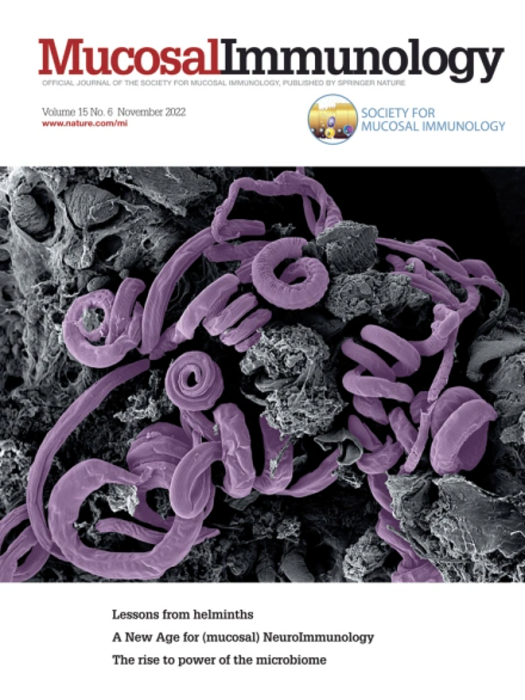
Mucosal Immunology
Mucosal Immunology 医学-免疫学
CiteScore
16.60
自引率
3.80%
发文量
100
审稿时长
12 days
期刊介绍: Mucosal Immunology, the official publication of the Society of Mucosal Immunology (SMI), serves as a forum for both basic and clinical scientists to discuss immunity and inflammation involving mucosal tissues. It covers gastrointestinal, pulmonary, nasopharyngeal, oral, ocular, and genitourinary immunology through original research articles, scholarly reviews, commentaries, editorials, and letters. The journal gives equal consideration to basic, translational, and clinical studies and also serves as a primary communication channel for the SMI governing board and its members, featuring society news, meeting announcements, policy discussions, and job/training opportunities advertisements.
×
引用
GB/T 7714-2015
复制
MLA
复制
APA
复制
导出至
BibTeX EndNote RefMan NoteFirst NoteExpress
×
提示
您的信息不完整,为了账户安全,请先补充。
现在去补充
×
提示
您因"违规操作"
具体请查看互助需知
我知道了
×
提示
确定
请完成安全验证×
copy
已复制链接
快去分享给好友吧!
我知道了
右上角分享
点击右上角分享
0
联系我们:info@booksci.cn Book学术提供免费学术资源搜索服务,方便国内外学者检索中英文文献。致力于提供最便捷和优质的服务体验。 Copyright © 2023 布克学术 All rights reserved.
京ICP备2023020795号-1
ghs 京公网安备 11010802042870号
Book学术文献互助
Book学术文献互助群
群 号:481959085
Book学术官方微信