An experimental system for detection and localization of hemorrhage using ultra-wideband microwaves with deep learning

Eisa Hedayati, Fatemeh Safari, George Verghese, Vito R. Ciancia, Daniel K. Sodickson, Seena Dehkharghani, Leeor Alon
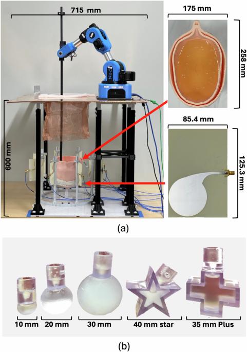
{"title":"An experimental system for detection and localization of hemorrhage using ultra-wideband microwaves with deep learning","authors":"Eisa Hedayati, Fatemeh Safari, George Verghese, Vito R. Ciancia, Daniel K. Sodickson, Seena Dehkharghani, Leeor Alon","doi":"10.1038/s44172-024-00259-4","DOIUrl":null,"url":null,"abstract":"Stroke is a leading cause of mortality and disability. Emergent diagnosis and intervention are critical, and predicated upon initial brain imaging; however, existing clinical imaging modalities are generally costly, immobile, and demand highly specialized operation and interpretation. Low-energy microwaves have been explored as a low-cost, small form factor, fast, and safe probe for tissue dielectric properties measurements, with both imaging and diagnostic potential. Nevertheless, challenges inherent to microwave reconstruction have impeded progress, hence conduction of microwave imaging remains an elusive scientific aim. Herein, we introduce a dedicated experimental framework comprising a robotic navigation system to translate blood-mimicking phantoms within a human head model. An 8-element ultra-wideband array of modified antipodal Vivaldi antennas was developed and driven by a two-port vector network analyzer spanning 0.6–9.0 GHz at an operating power of 1 mW. Complex scattering parameters were measured, and dielectric signatures of hemorrhage were learned using a dedicated deep neural network for prediction of hemorrhage classes and localization. An overall sensitivity and specificity for detection >0.99 was observed, with Rayleigh mean localization error of 1.65 mm. The study establishes the feasibility of a robust experimental model and deep learning solution for ultra-wideband microwave stroke detection. Eisa Hedayati, Fatemeh Safari and colleagues use an array of ultra-wideband microwave antennas to locate the haemorrhages in a human head phantom. The results of the measurements are processed by the deep neural network algorithm to classify the digital signatures for efficient detection and localization.","PeriodicalId":72644,"journal":{"name":"Communications engineering","volume":" ","pages":"1-10"},"PeriodicalIF":0.0000,"publicationDate":"2024-09-05","publicationTypes":"Journal Article","fieldsOfStudy":null,"isOpenAccess":false,"openAccessPdf":"https://www.nature.com/articles/s44172-024-00259-4.pdf","citationCount":"0","resultStr":null,"platform":"Semanticscholar","paperid":null,"PeriodicalName":"Communications engineering","FirstCategoryId":"1085","ListUrlMain":"https://www.nature.com/articles/s44172-024-00259-4","RegionNum":0,"RegionCategory":null,"ArticlePicture":[],"TitleCN":null,"AbstractTextCN":null,"PMCID":null,"EPubDate":"","PubModel":"","JCR":"","JCRName":"","Score":null,"Total":0}
引用次数: 0

Abstract

Stroke is a leading cause of mortality and disability. Emergent diagnosis and intervention are critical, and predicated upon initial brain imaging; however, existing clinical imaging modalities are generally costly, immobile, and demand highly specialized operation and interpretation. Low-energy microwaves have been explored as a low-cost, small form factor, fast, and safe probe for tissue dielectric properties measurements, with both imaging and diagnostic potential. Nevertheless, challenges inherent to microwave reconstruction have impeded progress, hence conduction of microwave imaging remains an elusive scientific aim. Herein, we introduce a dedicated experimental framework comprising a robotic navigation system to translate blood-mimicking phantoms within a human head model. An 8-element ultra-wideband array of modified antipodal Vivaldi antennas was developed and driven by a two-port vector network analyzer spanning 0.6–9.0 GHz at an operating power of 1 mW. Complex scattering parameters were measured, and dielectric signatures of hemorrhage were learned using a dedicated deep neural network for prediction of hemorrhage classes and localization. An overall sensitivity and specificity for detection >0.99 was observed, with Rayleigh mean localization error of 1.65 mm. The study establishes the feasibility of a robust experimental model and deep learning solution for ultra-wideband microwave stroke detection. Eisa Hedayati, Fatemeh Safari and colleagues use an array of ultra-wideband microwave antennas to locate the haemorrhages in a human head phantom. The results of the measurements are processed by the deep neural network algorithm to classify the digital signatures for efficient detection and localization.
Abstract Image

利用超宽带微波和深度学习检测和定位出血的实验系统
脑卒中是导致死亡和残疾的主要原因。紧急诊断和干预至关重要,并以初步脑成像为基础;然而,现有的临床成像模式通常成本高昂、无法移动,并且需要高度专业化的操作和解释。低能量微波作为一种低成本、小尺寸、快速和安全的探针,已被用于组织介电特性测量,具有成像和诊断潜力。然而,微波重建所固有的挑战阻碍了研究的进展,因此微波成像仍然是一个难以实现的科学目标。在本文中,我们介绍了一个专用的实验框架,该框架由一个机器人导航系统组成,用于在人体头部模型中转换仿血模型。我们开发了一个 8 元超宽带阵列,该阵列由改进的反脚维瓦尔第天线组成,并由双端口矢量网络分析仪驱动,工作功率为 1 mW,频率跨度为 0.6-9.0 GHz。测量了复杂的散射参数,并使用专用的深度神经网络学习了出血的介电特征,以预测出血类别和定位。总体检测灵敏度和特异性均为 0.99,瑞利平均定位误差为 1.65 毫米。该研究证明了超宽带微波中风检测的稳健实验模型和深度学习解决方案的可行性。Eisa Hedayati、Fatemeh Safari 及其同事使用超宽带微波天线阵列定位人体头部模型中的出血点。测量结果经过深度神经网络算法处理,对数字签名进行分类,从而实现高效检测和定位。
本文章由计算机程序翻译,如有差异,请以英文原文为准。
求助全文
约1分钟内获得全文 求助全文
来源期刊
自引率
0.00%
发文量
0
×
引用
GB/T 7714-2015
复制
MLA
复制
APA
复制
导出至
BibTeX EndNote RefMan NoteFirst NoteExpress
×
提示
您的信息不完整,为了账户安全,请先补充。
现在去补充
×
提示
您因"违规操作"
具体请查看互助需知
我知道了
×
提示
确定
请完成安全验证×
copy
已复制链接
快去分享给好友吧!
我知道了
右上角分享
点击右上角分享
0
联系我们:info@booksci.cn Book学术提供免费学术资源搜索服务,方便国内外学者检索中英文文献。致力于提供最便捷和优质的服务体验。 Copyright © 2023 布克学术 All rights reserved.
京ICP备2023020795号-1
ghs 京公网安备 11010802042870号
Book学术文献互助
Book学术文献互助群
群 号:481959085
Book学术官方微信