{"title":"Root volume measurements of maxillary canines and lateral incisors in patients with unilateral maxillary canine impaction.","authors":"Mostafa Shahabi, Hossein Hosseini Zarch, Zahra Shadman, Farzaneh Ahrari","doi":"10.1590/2177-6709.29.4.e242416.oar","DOIUrl":null,"url":null,"abstract":"<p><strong>Objective: </strong>This study aimed to assess root volumes of maxillary canines and adjacent lateral incisors in patients with unilateral maxillary canine impaction.</p><p><strong>Methods: </strong>This cross-sectional study was performed on cone-beam computed tomography (CBCT) scans of 100 patients (49 females and 51 males) with unilateral maxillary canine impaction. The images were loaded in Planmeca Romexis Viewer, and root layers between the cementoenamel junction and apex were reconstructed at 600-µm intervals. At each layer, the root boundary was marked, and finally, the root volume was calculated by multiplying the layers' area by the thickness of 600 µm. The root size of canines and lateral incisors was compared between the impaction and normal eruption sides.</p><p><strong>Results: </strong>Sixty-two patients showed buccal canine impaction, and 38 presented palatal impaction. The mean root volume of canines on the impaction side was significantly greater than that on the normal eruption side; either the tooth was buccally or palatally impacted (p<0.001). The lateral incisors on the side of buccally-impacted canines showed a significantly smaller root volume than that of the contralateral side (p<0.001). However, there was no significant difference in the root size of lateral incisors between the two sides in cases presenting palatal canine impaction (p=0.177).</p><p><strong>Conclusion: </strong>The difference in root volume of canines between the two sides can serve as an indicator of canine impaction. The reduction in the root size of the lateral incisor on the side of the buccally impacted canine may be due to root resorption created by pressure from the canine's crown.</p>","PeriodicalId":38720,"journal":{"name":"Dental Press Journal of Orthodontics","volume":"29 4","pages":"e242416"},"PeriodicalIF":0.0000,"publicationDate":"2024-09-02","publicationTypes":"Journal Article","fieldsOfStudy":null,"isOpenAccess":false,"openAccessPdf":"https://www.ncbi.nlm.nih.gov/pmc/articles/PMC11368238/pdf/","citationCount":"0","resultStr":null,"platform":"Semanticscholar","paperid":null,"PeriodicalName":"Dental Press Journal of Orthodontics","FirstCategoryId":"1085","ListUrlMain":"https://doi.org/10.1590/2177-6709.29.4.e242416.oar","RegionNum":0,"RegionCategory":null,"ArticlePicture":[],"TitleCN":null,"AbstractTextCN":null,"PMCID":null,"EPubDate":"2024/1/1 0:00:00","PubModel":"eCollection","JCR":"Q2","JCRName":"Medicine","Score":null,"Total":0}
引用次数: 0
Abstract
Objective: This study aimed to assess root volumes of maxillary canines and adjacent lateral incisors in patients with unilateral maxillary canine impaction.
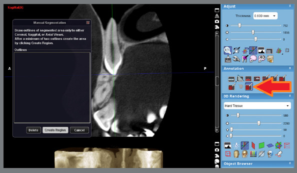
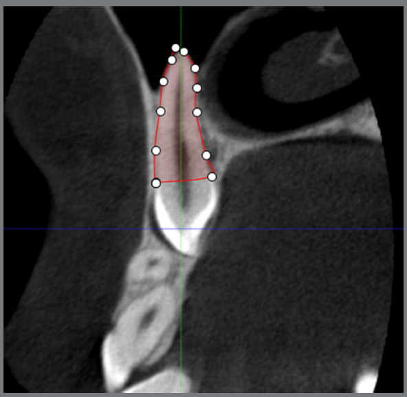
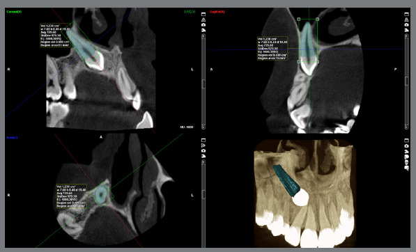
Methods: This cross-sectional study was performed on cone-beam computed tomography (CBCT) scans of 100 patients (49 females and 51 males) with unilateral maxillary canine impaction. The images were loaded in Planmeca Romexis Viewer, and root layers between the cementoenamel junction and apex were reconstructed at 600-µm intervals. At each layer, the root boundary was marked, and finally, the root volume was calculated by multiplying the layers' area by the thickness of 600 µm. The root size of canines and lateral incisors was compared between the impaction and normal eruption sides.
Results: Sixty-two patients showed buccal canine impaction, and 38 presented palatal impaction. The mean root volume of canines on the impaction side was significantly greater than that on the normal eruption side; either the tooth was buccally or palatally impacted (p<0.001). The lateral incisors on the side of buccally-impacted canines showed a significantly smaller root volume than that of the contralateral side (p<0.001). However, there was no significant difference in the root size of lateral incisors between the two sides in cases presenting palatal canine impaction (p=0.177).
Conclusion: The difference in root volume of canines between the two sides can serve as an indicator of canine impaction. The reduction in the root size of the lateral incisor on the side of the buccally impacted canine may be due to root resorption created by pressure from the canine's crown.
期刊介绍:
The Dental Press Journal of Orthodontics publishes scientific research articles, significant reviews, clinical and technical case reports, brief communications, and other materials related to Orthodontics and Facial Orthopedics.

 求助内容:
求助内容: 应助结果提醒方式:


