Quantitative microbiology with widefield microscopy: navigating optical artefacts for accurate interpretations

Georgeos Hardo, Ruizhe Li, Somenath Bakshi
{"title":"Quantitative microbiology with widefield microscopy: navigating optical artefacts for accurate interpretations","authors":"Georgeos Hardo, Ruizhe Li, Somenath Bakshi","doi":"10.1038/s44303-024-00024-4","DOIUrl":null,"url":null,"abstract":"Time-resolved live-cell imaging using widefield microscopy is instrumental in quantitative microbiology research. It allows researchers to track and measure the size, shape, and content of individual microbial cells over time. However, the small size of microbial cells poses a significant challenge in interpreting image data, as their dimensions approache that of the microscope’s depth of field, and they begin to experience significant diffraction effects. As a result, 2D widefield images of microbial cells contain projected 3D information, blurred by the 3D point spread function. In this study, we employed simulations and targeted experiments to investigate the impact of diffraction and projection on our ability to quantify the size and content of microbial cells from 2D microscopic images. This study points to some new and often unconsidered artefacts resulting from the interplay of projection and diffraction effects, within the context of quantitative microbiology. These artefacts introduce substantial errors and biases in size, fluorescence quantification, and even single-molecule counting, making the elimination of these errors a complex task. Awareness of these artefacts is crucial for designing strategies to accurately interpret micrographs of microbes. To address this, we present new experimental designs and machine learning-based analysis methods that account for these effects, resulting in accurate quantification of microbiological processes.","PeriodicalId":501709,"journal":{"name":"npj Imaging","volume":" ","pages":"1-16"},"PeriodicalIF":0.0000,"publicationDate":"2024-09-02","publicationTypes":"Journal Article","fieldsOfStudy":null,"isOpenAccess":false,"openAccessPdf":"https://www.nature.com/articles/s44303-024-00024-4.pdf","citationCount":"0","resultStr":null,"platform":"Semanticscholar","paperid":null,"PeriodicalName":"npj Imaging","FirstCategoryId":"1085","ListUrlMain":"https://www.nature.com/articles/s44303-024-00024-4","RegionNum":0,"RegionCategory":null,"ArticlePicture":[],"TitleCN":null,"AbstractTextCN":null,"PMCID":null,"EPubDate":"","PubModel":"","JCR":"","JCRName":"","Score":null,"Total":0}
引用次数: 0

Abstract

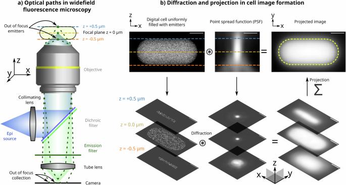
Time-resolved live-cell imaging using widefield microscopy is instrumental in quantitative microbiology research. It allows researchers to track and measure the size, shape, and content of individual microbial cells over time. However, the small size of microbial cells poses a significant challenge in interpreting image data, as their dimensions approache that of the microscope’s depth of field, and they begin to experience significant diffraction effects. As a result, 2D widefield images of microbial cells contain projected 3D information, blurred by the 3D point spread function. In this study, we employed simulations and targeted experiments to investigate the impact of diffraction and projection on our ability to quantify the size and content of microbial cells from 2D microscopic images. This study points to some new and often unconsidered artefacts resulting from the interplay of projection and diffraction effects, within the context of quantitative microbiology. These artefacts introduce substantial errors and biases in size, fluorescence quantification, and even single-molecule counting, making the elimination of these errors a complex task. Awareness of these artefacts is crucial for designing strategies to accurately interpret micrographs of microbes. To address this, we present new experimental designs and machine learning-based analysis methods that account for these effects, resulting in accurate quantification of microbiological processes.
Abstract Image

使用宽视野显微镜进行微生物定量分析:利用光学伪影进行准确解释
使用宽场显微镜进行时间分辨活细胞成像在定量微生物学研究中非常重要。研究人员可以利用它跟踪和测量单个微生物细胞随时间变化的大小、形状和内容。然而,由于微生物细胞的尺寸接近显微镜的景深,并且开始出现明显的衍射效应,因此细胞的小尺寸给解读图像数据带来了巨大挑战。因此,微生物细胞的二维宽视场图像包含投射的三维信息,而三维点扩散函数使其变得模糊不清。在这项研究中,我们利用模拟和针对性实验来研究衍射和投影对我们从二维显微图像量化微生物细胞大小和内容的能力的影响。这项研究指出,在定量微生物学中,投影和衍射效应的相互作用会产生一些新的、通常未被考虑的伪影。这些伪影在尺寸、荧光定量甚至单分子计数方面都会带来很大的误差和偏差,因此消除这些误差是一项复杂的任务。认识这些伪影对于设计准确解读微生物显微照片的策略至关重要。为了解决这个问题,我们提出了新的实验设计和基于机器学习的分析方法,以考虑这些影响,从而准确量化微生物过程。
本文章由计算机程序翻译,如有差异,请以英文原文为准。
求助全文
约1分钟内获得全文 求助全文
来源期刊
自引率
0.00%
发文量
0
×
引用
GB/T 7714-2015
复制
MLA
复制
APA
复制
导出至
BibTeX EndNote RefMan NoteFirst NoteExpress
×
提示
您的信息不完整,为了账户安全,请先补充。
现在去补充
×
提示
您因"违规操作"
具体请查看互助需知
我知道了
×
提示
确定
请完成安全验证×
copy
已复制链接
快去分享给好友吧!
我知道了
右上角分享
点击右上角分享
0
联系我们:info@booksci.cn Book学术提供免费学术资源搜索服务,方便国内外学者检索中英文文献。致力于提供最便捷和优质的服务体验。 Copyright © 2023 布克学术 All rights reserved.
京ICP备2023020795号-1
ghs 京公网安备 11010802042870号
Book学术文献互助
Book学术文献互助群
群 号:481959085
Book学术官方微信