Alexandre Bellier, P Tafforeau, A Bouziane, T Angelloz-Nicoud, P D Lee, C Walsh
{"title":"Micro to macro scale anatomical analysis of the human hippocampal arteries with synchrotron hierarchical phase-contrast tomography.","authors":"Alexandre Bellier, P Tafforeau, A Bouziane, T Angelloz-Nicoud, P D Lee, C Walsh","doi":"10.1007/s00276-024-03467-x","DOIUrl":null,"url":null,"abstract":"<p><strong>Purpose: </strong>To date, no non-invasive imaging modality has been employed to profile the structural intricacies of the hippocampal arterial microvasculature in humans. We hypothesised that synchrotron-based imaging of the human hippocampus would enable precise characterisation of the arterial microvasculature.</p><p><strong>Methods: </strong>Two preserved human brains from, a 69-year-old female and a 63-year-old male body donors were imaged using hierarchical phase-contrast tomography (HiP-CT) with synchrotron radiation at multiple voxel resolutions from 25.08 μm down to 2.45 μm. Subsequent manual and semi-automatic artery segmentation were performed followed by morphometric analyses. These data were compared to published data from alternative methodologies.</p><p><strong>Results: </strong>HiP-CT made it possible to segment in context the arterial architecture of the human hippocampus. Our analysis identified anterior, medial and posterior hippocampal arteries arising from the P2 segment of the posterior cerebral artery on the image slices. We mapped arterial branches with external diameters greater than 50 μm in the hippocampal region. We visualised vascular asymmetry and quantified arterial structures with diameters as small as 7 μm.</p><p><strong>Conclusions: </strong>Through the application of HiP-CT, we have provided the first imaging visualisation and quantification of the arterial system of the human hippocampus at high resolution in the context of whole brain imaging. Our results bridge the gap between anatomical and histological scales.</p>","PeriodicalId":49461,"journal":{"name":"Surgical and Radiologic Anatomy","volume":null,"pages":null},"PeriodicalIF":1.4000,"publicationDate":"2024-11-01","publicationTypes":"Journal Article","fieldsOfStudy":null,"isOpenAccess":false,"openAccessPdf":"https://www.ncbi.nlm.nih.gov/pmc/articles/PMC11458648/pdf/","citationCount":"0","resultStr":null,"platform":"Semanticscholar","paperid":null,"PeriodicalName":"Surgical and Radiologic Anatomy","FirstCategoryId":"3","ListUrlMain":"https://doi.org/10.1007/s00276-024-03467-x","RegionNum":4,"RegionCategory":"医学","ArticlePicture":[],"TitleCN":null,"AbstractTextCN":null,"PMCID":null,"EPubDate":"2024/9/3 0:00:00","PubModel":"Epub","JCR":"Q2","JCRName":"Medicine","Score":null,"Total":0}
引用次数: 0
Abstract
Purpose: To date, no non-invasive imaging modality has been employed to profile the structural intricacies of the hippocampal arterial microvasculature in humans. We hypothesised that synchrotron-based imaging of the human hippocampus would enable precise characterisation of the arterial microvasculature.
Methods: Two preserved human brains from, a 69-year-old female and a 63-year-old male body donors were imaged using hierarchical phase-contrast tomography (HiP-CT) with synchrotron radiation at multiple voxel resolutions from 25.08 μm down to 2.45 μm. Subsequent manual and semi-automatic artery segmentation were performed followed by morphometric analyses. These data were compared to published data from alternative methodologies.
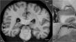
Results: HiP-CT made it possible to segment in context the arterial architecture of the human hippocampus. Our analysis identified anterior, medial and posterior hippocampal arteries arising from the P2 segment of the posterior cerebral artery on the image slices. We mapped arterial branches with external diameters greater than 50 μm in the hippocampal region. We visualised vascular asymmetry and quantified arterial structures with diameters as small as 7 μm.
Conclusions: Through the application of HiP-CT, we have provided the first imaging visualisation and quantification of the arterial system of the human hippocampus at high resolution in the context of whole brain imaging. Our results bridge the gap between anatomical and histological scales.
期刊介绍:
Anatomy is a morphological science which cannot fail to interest the clinician. The practical application of anatomical research to clinical problems necessitates special adaptation and selectivity in choosing from numerous international works. Although there is a tendency to believe that meaningful advances in anatomy are unlikely, constant revision is necessary. Surgical and Radiologic Anatomy, the first international journal of Clinical anatomy has been created in this spirit.
Its goal is to serve clinicians, regardless of speciality-physicians, surgeons, radiologists or other specialists-as an indispensable aid with which they can improve their knowledge of anatomy. Each issue includes: Original papers, review articles, articles on the anatomical bases of medical, surgical and radiological techniques, articles of normal radiologic anatomy, brief reviews of anatomical publications of clinical interest.
Particular attention is given to high quality illustrations, which are indispensable for a better understanding of anatomical problems.
Surgical and Radiologic Anatomy is a journal written by anatomists for clinicians with a special interest in anatomy.

 求助内容:
求助内容: 应助结果提醒方式:


