A parafoveal retinal cones analysis using adaptive-optics retinal camera in patients with primary open angle glaucoma
IF 2.8
3区 医学
Q1 OPHTHALMOLOGY
引用次数: 0
Abstract
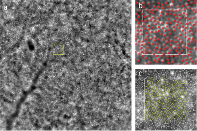
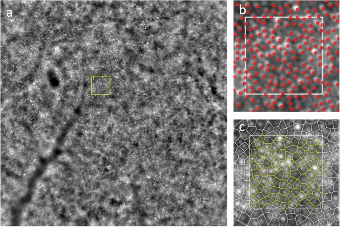
To study the density, spacing, and regularity of retinal cone photoreceptors using an Adaptive Optics (AO) retinal camera (Rtx1TM, Imagine Eyes, Orsay, France) in patients with Primary Open Angle Glaucoma (POAG) and to compare the outcomes with those of healthy age-matched control subjects. The study included 43 eyes with POAG and 31 eyes of normal subjects. POAG patients were divided into three groups according to the severity of the visual field defect. The AO Rtx1TM was used to obtain images of the parafoveal cone mosaic to calculate cone values. Analysis was performed at two and four degrees of eccentricity from the fovea along the four meridians (nasal, temporal, superior, inferior). In POAG eyes, the mean ± standard deviation (SD) cone density at 2° considering all meridians was significantly lower than in normal controls (23,058.6 ± 3532.0 cones/mm2, and 25,511.7 ± 3157.5 cones/mm2, respectively; p = 0.003). Cone spacing was 7.3 ± 0.5 µm in POAG and 7.0 ± 0.4 µm in normal controls (p = 0.005), and cone regularity was 90.5 ± 4.9% and 93.5 ± 1.9% in POAG and normal controls, respectively (p < 0.001). At 4° similar trends were observed. However, no significant differences were found among patients with different severity of POAG (p > 0.05). Using AO Rtx1TM, significant differences in retinal photoreceptors mosaic pattern were found between POAG eyes and age-matched controls, indicating a reduction in photoreceptors in POAG. No significant differences in retinal photoreceptor values were found among the three POAG groups.


利用自适应光学视网膜照相机对原发性开角型青光眼患者的眼底视锥进行分析。
研究目的使用自适应光学(AO)视网膜照相机(Rtx1TM,Imagine Eyes,法国奥赛公司)研究原发性开角型青光眼(POAG)患者视网膜视锥光感受器的密度、间距和规则性,并将结果与年龄匹配的健康对照组进行比较:研究对象包括 43 名 POAG 患者和 31 名正常人。根据视野缺损的严重程度,POAG 患者被分为三组。使用 AO Rtx1TM 获取视网膜旁视锥镶嵌图像,计算视锥值。沿四条经线(鼻经、颞经、上经、下经)在距离眼窝两度和四度偏心时进行分析:在 POAG 眼中,考虑到所有经线,偏心 2 度处的视锥密度平均值 ± 标准差 (SD) 明显低于正常对照组(分别为 23058.6 ± 3532.0 个视锥/平方毫米和 25511.7 ± 3157.5 个视锥/平方毫米;P = 0.003)。POAG 和正常对照组的锥体间距分别为 7.3 ± 0.5 µm 和 7.0 ± 0.4 µm (p = 0.005),锥体规整度分别为 90.5 ± 4.9% 和 93.5 ± 1.9% (p 0.05):结论:使用 AO Rtx1TM,POAG 眼睛与年龄匹配的对照组之间视网膜感光器镶嵌模式存在显著差异,表明 POAG 患者的感光器减少。在三个 POAG 组别中,视网膜光感受器值无明显差异。
本文章由计算机程序翻译,如有差异,请以英文原文为准。
求助全文
约1分钟内获得全文
求助全文
来源期刊

Eye
医学-眼科学
CiteScore
6.40
自引率
5.10%
发文量
481
审稿时长
3-6 weeks
期刊介绍:
Eye seeks to provide the international practising ophthalmologist with high quality articles, of academic rigour, on the latest global clinical and laboratory based research. Its core aim is to advance the science and practice of ophthalmology with the latest clinical- and scientific-based research. Whilst principally aimed at the practising clinician, the journal contains material of interest to a wider readership including optometrists, orthoptists, other health care professionals and research workers in all aspects of the field of visual science worldwide. Eye is the official journal of The Royal College of Ophthalmologists.
Eye encourages the submission of original articles covering all aspects of ophthalmology including: external eye disease; oculo-plastic surgery; orbital and lacrimal disease; ocular surface and corneal disorders; paediatric ophthalmology and strabismus; glaucoma; medical and surgical retina; neuro-ophthalmology; cataract and refractive surgery; ocular oncology; ophthalmic pathology; ophthalmic genetics.
 求助内容:
求助内容: 应助结果提醒方式:


