Katherine Miranda-Muñoz, Kirsten Midkiff, Alan Woessner, Mahyar Afshar-Mohajer, Min Zou, Erik Pollock, David Gonzalez-Nino, Gary Prinz, Lillian Hutchinson, Ruohan Li, Kushan Kompalage, Christopher T Culbertson, Ryan Jared Tucker, Hans Coetzee, Tsung Tsai, Jeremy Powell, Jorge Almodovar
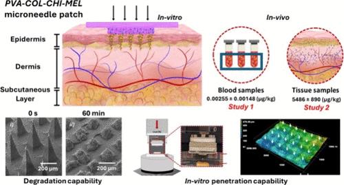
{"title":"A Multicomponent Microneedle Patch for the Delivery of Meloxicam for Veterinary Applications.","authors":"Katherine Miranda-Muñoz, Kirsten Midkiff, Alan Woessner, Mahyar Afshar-Mohajer, Min Zou, Erik Pollock, David Gonzalez-Nino, Gary Prinz, Lillian Hutchinson, Ruohan Li, Kushan Kompalage, Christopher T Culbertson, Ryan Jared Tucker, Hans Coetzee, Tsung Tsai, Jeremy Powell, Jorge Almodovar","doi":"10.1021/acsnano.4c08072","DOIUrl":null,"url":null,"abstract":"<p><p>This study evaluates the use of poly(vinyl alcohol), collagen, and chitosan blends for developing a microneedle patch for the delivery of meloxicam (MEL). Results confirm successful MEL encapsulation, structural integrity, and chemical stability even after ethylene oxide sterilization. Mechanical testing indicates the patch has the required properties for effective skin penetration and drug delivery, as demonstrated by load-displacement curves showing successful penetration of pig ear surfaces at 3N of normal load. <i>In vitro</i> imaging confirms the microneedle patch penetrates the pig's ear cadaver skin effectively and uniformly, with histological evaluation revealing the sustained presence and gradual degradation of microneedles within the skin. Additionally, <i>in vitro</i> drug diffusion experiments utilizing ballistic gel suggest that microneedles commence dissolution almost immediately upon insertion into the gel, steadily releasing the drug over 24 h. Furthermore, the microneedle patch demonstrates ideal drug release capabilities, achieving nearly 100% release of meloxicam content from a single patch within 18 h. Finally, <i>in vivo</i> studies using pigs demonstrate the successful dissolution and transdermal drug delivery efficacy of biodegradable microneedle patches delivering meloxicam in a porcine model, with over 70% of microneedles undergoing dissolution after 3 days. While low detectable meloxicam concentrations were observed in the bloodstream, high levels were detected in the ear tissue, confirming the release and diffusion of the drug from microneedles. This work highlights the potential of microneedle patches for controlled drug release in veterinary applications.</p>","PeriodicalId":21,"journal":{"name":"ACS Nano","volume":" ","pages":"25716-25739"},"PeriodicalIF":16.0000,"publicationDate":"2024-09-17","publicationTypes":"Journal Article","fieldsOfStudy":null,"isOpenAccess":false,"openAccessPdf":"","citationCount":"0","resultStr":null,"platform":"Semanticscholar","paperid":null,"PeriodicalName":"ACS Nano","FirstCategoryId":"88","ListUrlMain":"https://doi.org/10.1021/acsnano.4c08072","RegionNum":1,"RegionCategory":"材料科学","ArticlePicture":[],"TitleCN":null,"AbstractTextCN":null,"PMCID":null,"EPubDate":"2024/9/3 0:00:00","PubModel":"Epub","JCR":"Q1","JCRName":"CHEMISTRY, MULTIDISCIPLINARY","Score":null,"Total":0}
引用次数: 0
Abstract
This study evaluates the use of poly(vinyl alcohol), collagen, and chitosan blends for developing a microneedle patch for the delivery of meloxicam (MEL). Results confirm successful MEL encapsulation, structural integrity, and chemical stability even after ethylene oxide sterilization. Mechanical testing indicates the patch has the required properties for effective skin penetration and drug delivery, as demonstrated by load-displacement curves showing successful penetration of pig ear surfaces at 3N of normal load. In vitro imaging confirms the microneedle patch penetrates the pig's ear cadaver skin effectively and uniformly, with histological evaluation revealing the sustained presence and gradual degradation of microneedles within the skin. Additionally, in vitro drug diffusion experiments utilizing ballistic gel suggest that microneedles commence dissolution almost immediately upon insertion into the gel, steadily releasing the drug over 24 h. Furthermore, the microneedle patch demonstrates ideal drug release capabilities, achieving nearly 100% release of meloxicam content from a single patch within 18 h. Finally, in vivo studies using pigs demonstrate the successful dissolution and transdermal drug delivery efficacy of biodegradable microneedle patches delivering meloxicam in a porcine model, with over 70% of microneedles undergoing dissolution after 3 days. While low detectable meloxicam concentrations were observed in the bloodstream, high levels were detected in the ear tissue, confirming the release and diffusion of the drug from microneedles. This work highlights the potential of microneedle patches for controlled drug release in veterinary applications.
期刊介绍:
ACS Nano, published monthly, serves as an international forum for comprehensive articles on nanoscience and nanotechnology research at the intersections of chemistry, biology, materials science, physics, and engineering. The journal fosters communication among scientists in these communities, facilitating collaboration, new research opportunities, and advancements through discoveries. ACS Nano covers synthesis, assembly, characterization, theory, and simulation of nanostructures, nanobiotechnology, nanofabrication, methods and tools for nanoscience and nanotechnology, and self- and directed-assembly. Alongside original research articles, it offers thorough reviews, perspectives on cutting-edge research, and discussions envisioning the future of nanoscience and nanotechnology.
 求助内容:
求助内容: 应助结果提醒方式:


