{"title":"An unilateral double fenestration of the right external jugular vein: a rare variant.","authors":"R Dhivyaashree, Nandini Rajaram, Jahira Banu, Suman Verma, Hottigoudar Yekappa Suma","doi":"10.1007/s00276-024-03473-z","DOIUrl":null,"url":null,"abstract":"<p><strong>Objectives: </strong>This report presents a rare anatomical variation, double fenestration of the External jugular vein on the right side.</p><p><strong>Materials and methods: </strong>During the routine dissection of a male cadaver aged 60 years, we observed a unilateral large double fenestration of the External jugular vein on the right side.</p><p><strong>Results: </strong>After its formation from the posterior division of the retromandibular and posterior auricular veins, External jugular vein descended in the posterior triangle of neck. Here, it divided into medial, intermediate, and lateral veins that united again before draining into the subclavian vein. Lateral vein was the largest (7.2 cm) and intermediate and medial veins were measuring 6.4 cm each. Two large fenestrations, measuring 5.8 cm each, arranged like a \"double bubble\" were seen in the External jugular vein extending from fourth to sixth cervical (C4 to C6) vertebrae. The medial branch of supraclavicular nerve was seen passing superficial to the distal part of External jugular vein. On the left side, the course of External jugular vein showed a standard pattern.</p><p><strong>Conclusion: </strong>Surgeons must be acquainted with the varied anatomy of the superficial neck veins to prevent major bleeding during operative procedures, including carotid endarterectomy, flap operations, & central venous catheterisation.</p>","PeriodicalId":49461,"journal":{"name":"Surgical and Radiologic Anatomy","volume":" ","pages":"1807-1810"},"PeriodicalIF":1.4000,"publicationDate":"2024-11-01","publicationTypes":"Journal Article","fieldsOfStudy":null,"isOpenAccess":false,"openAccessPdf":"","citationCount":"0","resultStr":null,"platform":"Semanticscholar","paperid":null,"PeriodicalName":"Surgical and Radiologic Anatomy","FirstCategoryId":"3","ListUrlMain":"https://doi.org/10.1007/s00276-024-03473-z","RegionNum":4,"RegionCategory":"医学","ArticlePicture":[],"TitleCN":null,"AbstractTextCN":null,"PMCID":null,"EPubDate":"2024/8/30 0:00:00","PubModel":"Epub","JCR":"Q2","JCRName":"Medicine","Score":null,"Total":0}
引用次数: 0
Abstract
Objectives: This report presents a rare anatomical variation, double fenestration of the External jugular vein on the right side.
Materials and methods: During the routine dissection of a male cadaver aged 60 years, we observed a unilateral large double fenestration of the External jugular vein on the right side.
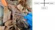
Results: After its formation from the posterior division of the retromandibular and posterior auricular veins, External jugular vein descended in the posterior triangle of neck. Here, it divided into medial, intermediate, and lateral veins that united again before draining into the subclavian vein. Lateral vein was the largest (7.2 cm) and intermediate and medial veins were measuring 6.4 cm each. Two large fenestrations, measuring 5.8 cm each, arranged like a "double bubble" were seen in the External jugular vein extending from fourth to sixth cervical (C4 to C6) vertebrae. The medial branch of supraclavicular nerve was seen passing superficial to the distal part of External jugular vein. On the left side, the course of External jugular vein showed a standard pattern.
Conclusion: Surgeons must be acquainted with the varied anatomy of the superficial neck veins to prevent major bleeding during operative procedures, including carotid endarterectomy, flap operations, & central venous catheterisation.
期刊介绍:
Anatomy is a morphological science which cannot fail to interest the clinician. The practical application of anatomical research to clinical problems necessitates special adaptation and selectivity in choosing from numerous international works. Although there is a tendency to believe that meaningful advances in anatomy are unlikely, constant revision is necessary. Surgical and Radiologic Anatomy, the first international journal of Clinical anatomy has been created in this spirit.
Its goal is to serve clinicians, regardless of speciality-physicians, surgeons, radiologists or other specialists-as an indispensable aid with which they can improve their knowledge of anatomy. Each issue includes: Original papers, review articles, articles on the anatomical bases of medical, surgical and radiological techniques, articles of normal radiologic anatomy, brief reviews of anatomical publications of clinical interest.
Particular attention is given to high quality illustrations, which are indispensable for a better understanding of anatomical problems.
Surgical and Radiologic Anatomy is a journal written by anatomists for clinicians with a special interest in anatomy.

 求助内容:
求助内容: 应助结果提醒方式:


