Nixi Xu, Chang Jiang, Zixian Chen, Zhenzhou Feng, Chun Jiang, Yuanwu Cao
{"title":"A patient with cervical ligamentum flavum haematoma: case report.","authors":"Nixi Xu, Chang Jiang, Zixian Chen, Zhenzhou Feng, Chun Jiang, Yuanwu Cao","doi":"10.1038/s41394-024-00679-6","DOIUrl":null,"url":null,"abstract":"<p><strong>Introduction: </strong>Ligamentum flavum haematoma (LFH) is an extremely rare entity, found mostly in the lumbar and thoracic ligamentum flavum and seldom in the cervical ligamentum flavum. Cervical LFH can cause paralysis in patients. We describe a case of LFH in the cervical spine that accepted surgical treatment.</p><p><strong>Case presentation: </strong>A 70-year-old man with incomplete spinal cord injury presented with sudden paralysis of his left limbs for 10 days and hemi-hypaesthesia below the level of the right clavicle. Magnetic resonance imaging (MRI) showed a space-occupying lesion in the left ligamentum flavum between the C4 and C5 laminae. The preliminary diagnoses were concluded to be incomplete spinal cord injury, spinal epidural lesions, and cervical spinal stenosis. After a posterior C3-C6 laminectomy with lateral mass screw instrumentation, the muscle strength and sensation recovered partially. The lesion was greyish black and located in the ligamentum flavum. A pathological examination identified it as a haematoma of the ligamentum flavum. The patient was discharged 15 days after the operation and commenced rehabilitation.</p><p><strong>Discussion: </strong>The LFH was mainly caused by slight trauma during gentle activities and contributed by many factors. MRI is an essential tool but pathological diagnosis is the gold standard. Most LFH patients can be treated surgically.</p>","PeriodicalId":22079,"journal":{"name":"Spinal Cord Series and Cases","volume":"10 1","pages":"65"},"PeriodicalIF":0.9000,"publicationDate":"2024-08-29","publicationTypes":"Journal Article","fieldsOfStudy":null,"isOpenAccess":false,"openAccessPdf":"https://www.ncbi.nlm.nih.gov/pmc/articles/PMC11362265/pdf/","citationCount":"0","resultStr":null,"platform":"Semanticscholar","paperid":null,"PeriodicalName":"Spinal Cord Series and Cases","FirstCategoryId":"1085","ListUrlMain":"https://doi.org/10.1038/s41394-024-00679-6","RegionNum":0,"RegionCategory":null,"ArticlePicture":[],"TitleCN":null,"AbstractTextCN":null,"PMCID":null,"EPubDate":"","PubModel":"","JCR":"Q4","JCRName":"CLINICAL NEUROLOGY","Score":null,"Total":0}
引用次数: 0
Abstract
Introduction: Ligamentum flavum haematoma (LFH) is an extremely rare entity, found mostly in the lumbar and thoracic ligamentum flavum and seldom in the cervical ligamentum flavum. Cervical LFH can cause paralysis in patients. We describe a case of LFH in the cervical spine that accepted surgical treatment.
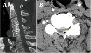
Case presentation: A 70-year-old man with incomplete spinal cord injury presented with sudden paralysis of his left limbs for 10 days and hemi-hypaesthesia below the level of the right clavicle. Magnetic resonance imaging (MRI) showed a space-occupying lesion in the left ligamentum flavum between the C4 and C5 laminae. The preliminary diagnoses were concluded to be incomplete spinal cord injury, spinal epidural lesions, and cervical spinal stenosis. After a posterior C3-C6 laminectomy with lateral mass screw instrumentation, the muscle strength and sensation recovered partially. The lesion was greyish black and located in the ligamentum flavum. A pathological examination identified it as a haematoma of the ligamentum flavum. The patient was discharged 15 days after the operation and commenced rehabilitation.
Discussion: The LFH was mainly caused by slight trauma during gentle activities and contributed by many factors. MRI is an essential tool but pathological diagnosis is the gold standard. Most LFH patients can be treated surgically.

 求助内容:
求助内容: 应助结果提醒方式:


