Semra Delibalta, Barış Genç, Meltem Ceyhan Bilgici, Kerim Aslan
{"title":"Feasibility study of computed high b-value diffusion-weighted magnetic resonance imaging for pediatric posterior fossa tumors.","authors":"Semra Delibalta, Barış Genç, Meltem Ceyhan Bilgici, Kerim Aslan","doi":"10.4274/dir.2024.242720","DOIUrl":null,"url":null,"abstract":"<p><strong>Purpose: </strong>To evaluate the diagnostic efficacy of computed diffusion-weighted imaging (DWI) in pediatric posterior fossa tumors generated using high b-values.</p><p><strong>Methods: </strong>We retrospectively performed our study on 32 pediatric patients who had undergone brain magnetic resonance imaging for a posterior fossa tumor between January 2016 and January 2022. The DWIs were evaluated for each patient by two blinded radiologists. The computed DWI (cDWI) was mathematically derived using a mono-exponential model from images with b = 0 and 1,000 s/mm<sup>2</sup> and high b-values of 1,500, 2,000, 3,000, and 5,000 s/mm<sup>2</sup>. The posterior fossa tumors were divided into two groups, low grade and high grade, and the tumor/thalamus signal intensity (SI) ratios were compared. The Mann-Whitney U test and receiver operating characteristic (ROC) curves were used to compare the diagnostic performance of the acquired DWI (DWI<sub>1000</sub>), apparent diffusion coefficient (ADC)<sub>1000</sub> maps, and cDWI (cDWI1500, cDWI<sub>2000</sub>, cDWI<sub>3000</sub>, and cDWI<sub>5000</sub>).</p><p><strong>Results: </strong>The comparison of the two tumor groups revealed that the tumor/thalamus SI ratio on the DWI<sub>1000</sub> and cDWI (cDWI1500, cDWI<sub>2000</sub>, cDWI<sub>3000</sub>, and cDWI<sub>5000</sub>) was statistically significantly higher in high-grade tumors (<i>P</i> < 0.001). In the ROC curve analysis, higher sensitivity and specificity were detected in the cDWI1500, cDWI<sub>2000</sub>, cDWI<sub>3000</sub>, and ADC<sub>1000</sub> maps (100%, 90.90%) compared with the DWI<sub>1000</sub> (80%, 81.80%). cDWI<sub>3000</sub> had the highest area under the curve (AUC) value compared with other parameters (AUC: 0.976).</p><p><strong>Conclusion: </strong>cDWI generated using high b-values was successful in differentiating between low-grade and high-grade posterior fossa tumors without increasing imaging time.</p><p><strong>Clinical significance: </strong>cDWI created using high b-values can provide additional information about tumor grade in pediatric posterior fossa tumors without requiring additional imaging time.</p>","PeriodicalId":11341,"journal":{"name":"Diagnostic and interventional radiology","volume":" ","pages":"526-531"},"PeriodicalIF":1.7000,"publicationDate":"2025-09-08","publicationTypes":"Journal Article","fieldsOfStudy":null,"isOpenAccess":false,"openAccessPdf":"https://www.ncbi.nlm.nih.gov/pmc/articles/PMC12417922/pdf/","citationCount":"0","resultStr":null,"platform":"Semanticscholar","paperid":null,"PeriodicalName":"Diagnostic and interventional radiology","FirstCategoryId":"3","ListUrlMain":"https://doi.org/10.4274/dir.2024.242720","RegionNum":4,"RegionCategory":"医学","ArticlePicture":[],"TitleCN":null,"AbstractTextCN":null,"PMCID":null,"EPubDate":"2024/9/2 0:00:00","PubModel":"Epub","JCR":"Q3","JCRName":"RADIOLOGY, NUCLEAR MEDICINE & MEDICAL IMAGING","Score":null,"Total":0}
引用次数: 0
Abstract
Purpose: To evaluate the diagnostic efficacy of computed diffusion-weighted imaging (DWI) in pediatric posterior fossa tumors generated using high b-values.
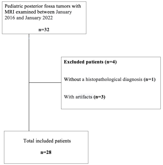
Methods: We retrospectively performed our study on 32 pediatric patients who had undergone brain magnetic resonance imaging for a posterior fossa tumor between January 2016 and January 2022. The DWIs were evaluated for each patient by two blinded radiologists. The computed DWI (cDWI) was mathematically derived using a mono-exponential model from images with b = 0 and 1,000 s/mm2 and high b-values of 1,500, 2,000, 3,000, and 5,000 s/mm2. The posterior fossa tumors were divided into two groups, low grade and high grade, and the tumor/thalamus signal intensity (SI) ratios were compared. The Mann-Whitney U test and receiver operating characteristic (ROC) curves were used to compare the diagnostic performance of the acquired DWI (DWI1000), apparent diffusion coefficient (ADC)1000 maps, and cDWI (cDWI1500, cDWI2000, cDWI3000, and cDWI5000).
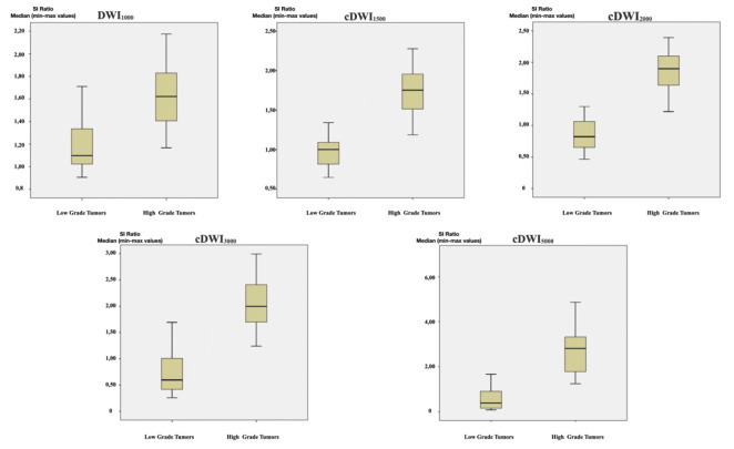
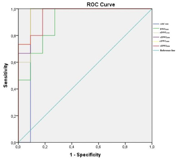
Results: The comparison of the two tumor groups revealed that the tumor/thalamus SI ratio on the DWI1000 and cDWI (cDWI1500, cDWI2000, cDWI3000, and cDWI5000) was statistically significantly higher in high-grade tumors (P < 0.001). In the ROC curve analysis, higher sensitivity and specificity were detected in the cDWI1500, cDWI2000, cDWI3000, and ADC1000 maps (100%, 90.90%) compared with the DWI1000 (80%, 81.80%). cDWI3000 had the highest area under the curve (AUC) value compared with other parameters (AUC: 0.976).
Conclusion: cDWI generated using high b-values was successful in differentiating between low-grade and high-grade posterior fossa tumors without increasing imaging time.
Clinical significance: cDWI created using high b-values can provide additional information about tumor grade in pediatric posterior fossa tumors without requiring additional imaging time.
期刊介绍:
Diagnostic and Interventional Radiology (Diagn Interv Radiol) is the open access, online-only official publication of Turkish Society of Radiology. It is published bimonthly and the journal’s publication language is English.
The journal is a medium for original articles, reviews, pictorial essays, technical notes related to all fields of diagnostic and interventional radiology.

 求助内容:
求助内容: 应助结果提醒方式:


