{"title":"Fluorescence and colorimetric analysis of β-estradiol based on aptamer assembled spherical nucleic acids†","authors":"Leyuan Chen, Aijiao Yuan, Dapeng Zhang, Wenjing Xie and Hanyong Peng","doi":"10.1039/D4AY01283F","DOIUrl":null,"url":null,"abstract":"<p >Detecting β-estradiol (E2) in environmental monitoring is a complex task due to its status as a significant environmental contaminant. The detection methods require precision, sensitivity, and the ability to be conducted on-site without expensive instrumentation. Herein, we developed a novel approach using E2 aptamer assembled spherical nucleic acids (SNAs), which combines the sensitivity of fluorescence and the simplicity of colorimetry. Initially, a fluorescein (FAM)-labeled DNA aptamer is attached to the surface of gold nanoparticles (AuNPs) through hybridization with thiol-labeled DNA, resulting in fluorescence quenching. However, when E2 is present, the aptamer specifically binds to it, displacing from the thiol-DNA and releasing from the AuNP's surface. This leads to the recovery of fluorescence, allowing for quantitative detection of E2 by measuring the increase in fluorescence signal. Additionally, E2 detection can also be achieved visually using ultraviolet light. For colorimetric analysis, we introduce another set of AuNPs modified with thiol-DNA complementary to the DNA remaining on the surface of the previous AuNPs. When E2 triggers the release of the aptamer, the DNA on both AuNPs hybridized to each other, causing the aggregation of AuNPs and resulting in a distinct color change from red to purple. The detection limits for fluorescence and colorimetric analyses are 1 nM and 5 nM, respectively. We successfully applied this biosensing strategy to determine E2 concentrations in tap water and serum samples. Furthermore, our assay exhibits high selectivity towards E2 over other estrogens. Overall, this innovative approach provides an effective and versatile method for convenient on-site monitoring of E2.</p>","PeriodicalId":64,"journal":{"name":"Analytical Methods","volume":null,"pages":null},"PeriodicalIF":2.7000,"publicationDate":"2024-08-26","publicationTypes":"Journal Article","fieldsOfStudy":null,"isOpenAccess":false,"openAccessPdf":"https://pubs.rsc.org/en/content/articlepdf/2024/ay/d4ay01283f?page=search","citationCount":"0","resultStr":null,"platform":"Semanticscholar","paperid":null,"PeriodicalName":"Analytical Methods","FirstCategoryId":"92","ListUrlMain":"https://pubs.rsc.org/en/content/articlelanding/2024/ay/d4ay01283f","RegionNum":3,"RegionCategory":"化学","ArticlePicture":[],"TitleCN":null,"AbstractTextCN":null,"PMCID":null,"EPubDate":"","PubModel":"","JCR":"Q2","JCRName":"CHEMISTRY, ANALYTICAL","Score":null,"Total":0}
引用次数: 0
Abstract
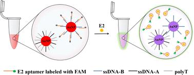
Detecting β-estradiol (E2) in environmental monitoring is a complex task due to its status as a significant environmental contaminant. The detection methods require precision, sensitivity, and the ability to be conducted on-site without expensive instrumentation. Herein, we developed a novel approach using E2 aptamer assembled spherical nucleic acids (SNAs), which combines the sensitivity of fluorescence and the simplicity of colorimetry. Initially, a fluorescein (FAM)-labeled DNA aptamer is attached to the surface of gold nanoparticles (AuNPs) through hybridization with thiol-labeled DNA, resulting in fluorescence quenching. However, when E2 is present, the aptamer specifically binds to it, displacing from the thiol-DNA and releasing from the AuNP's surface. This leads to the recovery of fluorescence, allowing for quantitative detection of E2 by measuring the increase in fluorescence signal. Additionally, E2 detection can also be achieved visually using ultraviolet light. For colorimetric analysis, we introduce another set of AuNPs modified with thiol-DNA complementary to the DNA remaining on the surface of the previous AuNPs. When E2 triggers the release of the aptamer, the DNA on both AuNPs hybridized to each other, causing the aggregation of AuNPs and resulting in a distinct color change from red to purple. The detection limits for fluorescence and colorimetric analyses are 1 nM and 5 nM, respectively. We successfully applied this biosensing strategy to determine E2 concentrations in tap water and serum samples. Furthermore, our assay exhibits high selectivity towards E2 over other estrogens. Overall, this innovative approach provides an effective and versatile method for convenient on-site monitoring of E2.
 求助内容:
求助内容: 应助结果提醒方式:


