Migration of an intrauterine contraceptive device into the bladder complicated by stone formation an exceptional complication: case report and literature review.
{"title":"Migration of an intrauterine contraceptive device into the bladder complicated by stone formation an exceptional complication: case report and literature review.","authors":"Hanane Houmaid, Karam Harou, Bouchra Fakhir, Ahlam Bassir, Lahcen Boukhanni, Abderrahim Aboulfalah, Hamid Asmouki, Abderraouf Soummani","doi":"10.1186/s40834-024-00302-x","DOIUrl":null,"url":null,"abstract":"<p><strong>Background: </strong>We report a rare and unusual case of intravesical migration of an intrauterine device with stone formation. The intrauterine device (IUD) is the most common method of reversible contraception in women. However, its insertion is not without risk, it can cause early or late complications. IUD can perforate the uterus wall and migrate into adjacent structures.</p><p><strong>Case presentation: </strong>A 35 year-old female 5 gravid, 4 para has been benefited from intrauterine contraceptive device (IUCD) 5 years ago, she was presented to gynecological consultation for chronic pelvic pain with urinary symptoms. There was history of a good IUD insertion 5 years ago, considered expelled after one month of its pose. Physical examination was normal, but a pelvic ultrasound and a plain abdominal radiography allowed the detection of an IUD outside the uterine cavity, but inside bladder. A diagnostic and therapeutic cystoscopy was performed, and the IUD with calculus was successfully removed. There were no postoperative complications.</p><p><strong>Conclusion: </strong>This case is reported to highlight and to reiterate the need to think about one of the rare complication of IUD insertion, which every practitioner must know, it's the transuterovesical migration, before concluding wrongly to its expulsion. It's a consequence of, non-compliance with the rules for inserting an IUD and poor monitoring. The evolution towards calcification is a certain consequence; its screening involves rigorous clinical monitoring.</p>","PeriodicalId":93956,"journal":{"name":"Contraception and reproductive medicine","volume":"9 1","pages":"42"},"PeriodicalIF":1.9000,"publicationDate":"2024-08-28","publicationTypes":"Journal Article","fieldsOfStudy":null,"isOpenAccess":false,"openAccessPdf":"https://www.ncbi.nlm.nih.gov/pmc/articles/PMC11351088/pdf/","citationCount":"0","resultStr":null,"platform":"Semanticscholar","paperid":null,"PeriodicalName":"Contraception and reproductive medicine","FirstCategoryId":"1085","ListUrlMain":"https://doi.org/10.1186/s40834-024-00302-x","RegionNum":0,"RegionCategory":null,"ArticlePicture":[],"TitleCN":null,"AbstractTextCN":null,"PMCID":null,"EPubDate":"","PubModel":"","JCR":"Q2","JCRName":"OBSTETRICS & GYNECOLOGY","Score":null,"Total":0}
引用次数: 0
Abstract
Background: We report a rare and unusual case of intravesical migration of an intrauterine device with stone formation. The intrauterine device (IUD) is the most common method of reversible contraception in women. However, its insertion is not without risk, it can cause early or late complications. IUD can perforate the uterus wall and migrate into adjacent structures.
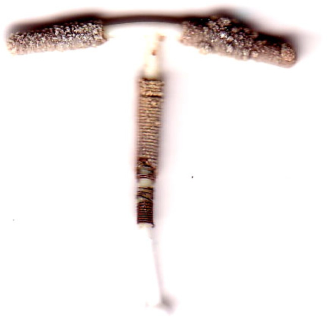
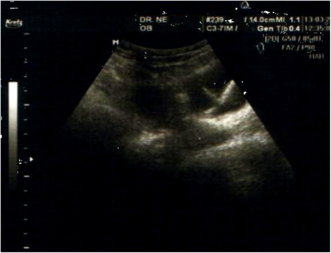
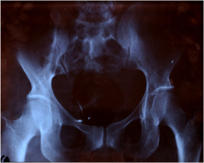
Case presentation: A 35 year-old female 5 gravid, 4 para has been benefited from intrauterine contraceptive device (IUCD) 5 years ago, she was presented to gynecological consultation for chronic pelvic pain with urinary symptoms. There was history of a good IUD insertion 5 years ago, considered expelled after one month of its pose. Physical examination was normal, but a pelvic ultrasound and a plain abdominal radiography allowed the detection of an IUD outside the uterine cavity, but inside bladder. A diagnostic and therapeutic cystoscopy was performed, and the IUD with calculus was successfully removed. There were no postoperative complications.
Conclusion: This case is reported to highlight and to reiterate the need to think about one of the rare complication of IUD insertion, which every practitioner must know, it's the transuterovesical migration, before concluding wrongly to its expulsion. It's a consequence of, non-compliance with the rules for inserting an IUD and poor monitoring. The evolution towards calcification is a certain consequence; its screening involves rigorous clinical monitoring.

 求助内容:
求助内容: 应助结果提醒方式:


