Ghassen Gader, Mohamed Amine Gharbi, Mohamed Ali Kharrat, Ahmed Harbaoui, Ihsèn Zammel
{"title":"Solitary thoracic spine osteochondroma: a rare cause for spinal cord compression.","authors":"Ghassen Gader, Mohamed Amine Gharbi, Mohamed Ali Kharrat, Ahmed Harbaoui, Ihsèn Zammel","doi":"10.1038/s41394-024-00677-8","DOIUrl":null,"url":null,"abstract":"<p><strong>Introduction: </strong>Osteochondromas, also known as osteocartilaginous exostosis, are among the most common benign cartilaginous bone tumors, primarily occurring as solitary lesions. While typically found in long bones, spinal involvement is rare, accounting for only a small percentage of benign lesions in this location. Solitary osteochondromas responsible for spinal cord compression are seldom.</p><p><strong>Case presentation: </strong>We describe the case of a 34-year-old male with no significant medical history, presenting with progressive symptoms suggestive of spinal cord compression. Imaging studies revealed a bony lesion originating from the left lateral aspect of the posterior arch of the T8 vertebra, causing spinal cord compression and myelopathy. Surgical intervention was necessary to decompress the spinal cord and obtain histological samples, resulting in immediate postoperative improvement in motor function. Pathologic exam concluded to an osteochondroma.</p><p><strong>Discussion: </strong>Osteochondromas primarily affect growing bones and are more commonly observed as solitary lesions, particularly in male patients. Spinal involvement is rare, and neurological symptoms are typically indicative of intracanalar extension of the exostosis, leading to compression of neural elements. Imaging modalities such as MRI are crucial for assessing cartilage thickness and the impact of compression on the spinal cord.</p>","PeriodicalId":22079,"journal":{"name":"Spinal Cord Series and Cases","volume":"10 1","pages":"63"},"PeriodicalIF":0.9000,"publicationDate":"2024-08-22","publicationTypes":"Journal Article","fieldsOfStudy":null,"isOpenAccess":false,"openAccessPdf":"https://www.ncbi.nlm.nih.gov/pmc/articles/PMC11341696/pdf/","citationCount":"0","resultStr":null,"platform":"Semanticscholar","paperid":null,"PeriodicalName":"Spinal Cord Series and Cases","FirstCategoryId":"1085","ListUrlMain":"https://doi.org/10.1038/s41394-024-00677-8","RegionNum":0,"RegionCategory":null,"ArticlePicture":[],"TitleCN":null,"AbstractTextCN":null,"PMCID":null,"EPubDate":"","PubModel":"","JCR":"Q4","JCRName":"CLINICAL NEUROLOGY","Score":null,"Total":0}
引用次数: 0
Abstract
Introduction: Osteochondromas, also known as osteocartilaginous exostosis, are among the most common benign cartilaginous bone tumors, primarily occurring as solitary lesions. While typically found in long bones, spinal involvement is rare, accounting for only a small percentage of benign lesions in this location. Solitary osteochondromas responsible for spinal cord compression are seldom.
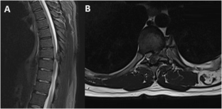
Case presentation: We describe the case of a 34-year-old male with no significant medical history, presenting with progressive symptoms suggestive of spinal cord compression. Imaging studies revealed a bony lesion originating from the left lateral aspect of the posterior arch of the T8 vertebra, causing spinal cord compression and myelopathy. Surgical intervention was necessary to decompress the spinal cord and obtain histological samples, resulting in immediate postoperative improvement in motor function. Pathologic exam concluded to an osteochondroma.
Discussion: Osteochondromas primarily affect growing bones and are more commonly observed as solitary lesions, particularly in male patients. Spinal involvement is rare, and neurological symptoms are typically indicative of intracanalar extension of the exostosis, leading to compression of neural elements. Imaging modalities such as MRI are crucial for assessing cartilage thickness and the impact of compression on the spinal cord.

 求助内容:
求助内容: 应助结果提醒方式:


