{"title":"Mixed hepatocellular carcinoma and high-grade neuroendocrine neoplasm with ambiguous histopathological features: a case report.","authors":"Kentaro Tsuji, Makoto Abe, Saho Wakamatsu, Sayuri Hoshi, Nobuo Hoshi, Chisato Takagi, Noriyoshi Fukushima, Kaoru Hirabayashi","doi":"10.1007/s00795-024-00396-x","DOIUrl":null,"url":null,"abstract":"<p><p>Well-differentiated neuroendocrine tumor (NET) and poorly differentiated neuroendocrine carcinoma (NEC) are distinct entities with different biological behavior. However, difficult cases showing equivocal morphology have been reported in some organs. Herein, we report a case of primary hepatic neuroendocrine neoplasm (NEN) with ambiguous histopathological features admixed with conventional hepatocellular carcinoma (HCC). A 70-year-old man with untreated chronic hepatitis B underwent left medial sectionectomy because of two incidental liver masses. On pathological examination, one of the resected tumors had intermingling NEN and HCC components. The NEN component consisted of relatively uniform tumor cells proliferating in trabecular, cord-like, or solid patterns with peripheral nuclear palisading. The tumor cells were immunopositive for synaptophysin, chromogranin A, cluster of differentiation 56 (CD56), and focally hepatocyte paraffin 1. p53 showed wild-type expression. The Ki-67 labeling index was 27% at the hot spot. Eleven months after the surgery, he died of a cerebral hemorrhage without evidence of recurrent liver cancer. The intermediate degree of differentiation and the modest proliferative activity can challenge the distinction between NEC and NET G3. While the coexisting HCC indicates NEC rather than NET in a pathogenetic viewpoint, such ambiguous tumor may not be as aggressive as typical NECs.</p>","PeriodicalId":18338,"journal":{"name":"Medical Molecular Morphology","volume":" ","pages":"62-68"},"PeriodicalIF":1.1000,"publicationDate":"2025-03-01","publicationTypes":"Journal Article","fieldsOfStudy":null,"isOpenAccess":false,"openAccessPdf":"","citationCount":"0","resultStr":null,"platform":"Semanticscholar","paperid":null,"PeriodicalName":"Medical Molecular Morphology","FirstCategoryId":"3","ListUrlMain":"https://doi.org/10.1007/s00795-024-00396-x","RegionNum":4,"RegionCategory":"医学","ArticlePicture":[],"TitleCN":null,"AbstractTextCN":null,"PMCID":null,"EPubDate":"2024/8/23 0:00:00","PubModel":"Epub","JCR":"Q3","JCRName":"PATHOLOGY","Score":null,"Total":0}
引用次数: 0
Abstract
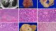
Well-differentiated neuroendocrine tumor (NET) and poorly differentiated neuroendocrine carcinoma (NEC) are distinct entities with different biological behavior. However, difficult cases showing equivocal morphology have been reported in some organs. Herein, we report a case of primary hepatic neuroendocrine neoplasm (NEN) with ambiguous histopathological features admixed with conventional hepatocellular carcinoma (HCC). A 70-year-old man with untreated chronic hepatitis B underwent left medial sectionectomy because of two incidental liver masses. On pathological examination, one of the resected tumors had intermingling NEN and HCC components. The NEN component consisted of relatively uniform tumor cells proliferating in trabecular, cord-like, or solid patterns with peripheral nuclear palisading. The tumor cells were immunopositive for synaptophysin, chromogranin A, cluster of differentiation 56 (CD56), and focally hepatocyte paraffin 1. p53 showed wild-type expression. The Ki-67 labeling index was 27% at the hot spot. Eleven months after the surgery, he died of a cerebral hemorrhage without evidence of recurrent liver cancer. The intermediate degree of differentiation and the modest proliferative activity can challenge the distinction between NEC and NET G3. While the coexisting HCC indicates NEC rather than NET in a pathogenetic viewpoint, such ambiguous tumor may not be as aggressive as typical NECs.
期刊介绍:
Medical Molecular Morphology is an international forum for researchers in both basic and clinical medicine to present and discuss new research on the structural mechanisms and the processes of health and disease at the molecular level. The structures of molecules, organelles, cells, tissues, and organs determine their normal function. Disease is thus best understood in terms of structural changes in these different levels of biological organization, especially in molecules and molecular interactions as well as the cellular localization of chemical components. Medical Molecular Morphology welcomes articles on basic or clinical research in the fields of cell biology, molecular biology, and medical, veterinary, and dental sciences using techniques for structural research such as electron microscopy, confocal laser scanning microscopy, enzyme histochemistry, immunohistochemistry, radioautography, X-ray microanalysis, and in situ hybridization.
Manuscripts submitted for publication must contain a statement to the effect that all human studies have been reviewed by the appropriate ethics committee and have therefore been performed in accordance with the ethical standards laid down in an appropriate version of the 1964 Declaration of Helsinki. It should also be stated clearly in the text that all persons gave their informed consent prior to their inclusion in the study. Details that might disclose the identity of the subjects under study should be omitted.
 求助内容:
求助内容: 应助结果提醒方式:


