{"title":"Muscle-derived IL-1β regulates EcSOD expression via the NBR1-p62-Nrf2 pathway in muscle during cancer cachexia","authors":"Mami Yamada, Eiji Warabi, Hisashi Oishi, Vitor A. Lira, Mitsuharu Okutsu","doi":"10.1113/JP286460","DOIUrl":null,"url":null,"abstract":"<div>\n \n <section>\n \n \n <div>Oxidative stress contributes to the loss of skeletal muscle mass and function in cancer cachexia. However, this outcome may be mitigated by an improved endogenous antioxidant defence system. Here, using the well-established oxidative stress-inducing muscle atrophy model of Lewis lung carcinoma (LLC) in 13-week-old male C57BL/6J mice, we demonstrate that extracellular superoxide dismutase (EcSOD) levels increase in the cachexia-prone extensor digitorum longus muscle. LLC transplantation significantly increased interleukin-1β (IL-1β) expression and release from extensor digitorum longus muscle fibres. Moreover, IL-1β treatment of C2C12 myotubes increased NBR1, p62 phosphorylation at Ser351, Nrf2 nuclear translocation and EcSOD protein expression. Additional studies <i>in vivo</i> indicated that intramuscular IL-1β injection is sufficient to stimulate EcSOD expression, which is prevented by muscle-specific knockout of p62 and Nrf2 (i.e. in p62 skmKO and Nrf2 skmKO mice, respectively). Finally, since an increase in circulating IL-1β may lead to unwanted outcomes, we demonstrate that targeting this pathway at p62 is sufficient to drive muscle EcSOD expression in an Nrf2-dependent manner. In summary, cancer cachexia increases EcSOD expression in extensor digitorum longus muscle via muscle-derived IL-1β-induced upregulation of p62 phosphorylation and Nrf2 activation. These findings provide further mechanistic evidence for the therapeutic potential of p62 and Nrf2 to mitigate cancer cachexia-induced muscle atrophy.\n\n <figure>\n <div><picture>\n <source></source></picture><p></p>\n </div>\n </figure>\n </div>\n </section>\n \n <section>\n \n <h3> Key points</h3>\n \n <div>\n <ul>\n \n <li>Oxidative stress plays an important role in muscle atrophy during cancer cachexia.</li>\n \n <li>EcSOD, which mitigates muscle loss during oxidative stress, is upregulated in 13-week-old male C57BL/6J mice of extensor digitorum longus muscles during cancer cachexia.</li>\n \n <li>Using mouse and cellular models, we demonstrate that cancer cachexia promotes muscle EcSOD protein expression via muscle-derived IL-1β-dependent stimulation of the NBR1-p62-Nrf2 signalling pathway.</li>\n \n <li>These results provide further evidence for the potential therapeutic targeting of the NBR1-p62-Nrf2 signalling pathway downstream of IL-1β to mitigate cancer cachexia-induced muscle atrophy.</li>\n </ul>\n </div>\n </section>\n </div>","PeriodicalId":50088,"journal":{"name":"Journal of Physiology-London","volume":"602 17","pages":"4215-4235"},"PeriodicalIF":4.7000,"publicationDate":"2024-08-21","publicationTypes":"Journal Article","fieldsOfStudy":null,"isOpenAccess":false,"openAccessPdf":"","citationCount":"0","resultStr":null,"platform":"Semanticscholar","paperid":null,"PeriodicalName":"Journal of Physiology-London","FirstCategoryId":"3","ListUrlMain":"https://onlinelibrary.wiley.com/doi/10.1113/JP286460","RegionNum":2,"RegionCategory":"医学","ArticlePicture":[],"TitleCN":null,"AbstractTextCN":null,"PMCID":null,"EPubDate":"","PubModel":"","JCR":"Q1","JCRName":"NEUROSCIENCES","Score":null,"Total":0}
引用次数: 0
Abstract
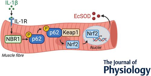
Oxidative stress contributes to the loss of skeletal muscle mass and function in cancer cachexia. However, this outcome may be mitigated by an improved endogenous antioxidant defence system. Here, using the well-established oxidative stress-inducing muscle atrophy model of Lewis lung carcinoma (LLC) in 13-week-old male C57BL/6J mice, we demonstrate that extracellular superoxide dismutase (EcSOD) levels increase in the cachexia-prone extensor digitorum longus muscle. LLC transplantation significantly increased interleukin-1β (IL-1β) expression and release from extensor digitorum longus muscle fibres. Moreover, IL-1β treatment of C2C12 myotubes increased NBR1, p62 phosphorylation at Ser351, Nrf2 nuclear translocation and EcSOD protein expression. Additional studies in vivo indicated that intramuscular IL-1β injection is sufficient to stimulate EcSOD expression, which is prevented by muscle-specific knockout of p62 and Nrf2 (i.e. in p62 skmKO and Nrf2 skmKO mice, respectively). Finally, since an increase in circulating IL-1β may lead to unwanted outcomes, we demonstrate that targeting this pathway at p62 is sufficient to drive muscle EcSOD expression in an Nrf2-dependent manner. In summary, cancer cachexia increases EcSOD expression in extensor digitorum longus muscle via muscle-derived IL-1β-induced upregulation of p62 phosphorylation and Nrf2 activation. These findings provide further mechanistic evidence for the therapeutic potential of p62 and Nrf2 to mitigate cancer cachexia-induced muscle atrophy.
Key points
Oxidative stress plays an important role in muscle atrophy during cancer cachexia.
EcSOD, which mitigates muscle loss during oxidative stress, is upregulated in 13-week-old male C57BL/6J mice of extensor digitorum longus muscles during cancer cachexia.
Using mouse and cellular models, we demonstrate that cancer cachexia promotes muscle EcSOD protein expression via muscle-derived IL-1β-dependent stimulation of the NBR1-p62-Nrf2 signalling pathway.
These results provide further evidence for the potential therapeutic targeting of the NBR1-p62-Nrf2 signalling pathway downstream of IL-1β to mitigate cancer cachexia-induced muscle atrophy.
期刊介绍:
The Journal of Physiology publishes full-length original Research Papers and Techniques for Physiology, which are short papers aimed at disseminating new techniques for physiological research. Articles solicited by the Editorial Board include Perspectives, Symposium Reports and Topical Reviews, which highlight areas of special physiological interest. CrossTalk articles are short editorial-style invited articles framing a debate between experts in the field on controversial topics. Letters to the Editor and Journal Club articles are also published. All categories of papers are subjected to peer reivew.
The Journal of Physiology welcomes submitted research papers in all areas of physiology. Authors should present original work that illustrates new physiological principles or mechanisms. Papers on work at the molecular level, at the level of the cell membrane, single cells, tissues or organs and on systems physiology are all acceptable. Theoretical papers and papers that use computational models to further our understanding of physiological processes will be considered if based on experimentally derived data and if the hypothesis advanced is directly amenable to experimental testing. While emphasis is on human and mammalian physiology, work on lower vertebrate or invertebrate preparations may be suitable if it furthers the understanding of the functioning of other organisms including mammals.
 求助内容:
求助内容: 应助结果提醒方式:


