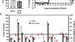
{"title":"Changes in cerebral vascular reactivity following mild repetitive head injury in awake rats: modeling the human experience.","authors":"Nicole Bens, Praveen Kulkarni, Craig F Ferris","doi":"10.1007/s00221-024-06907-7","DOIUrl":null,"url":null,"abstract":"<p><p>The changes in brain function in response to mild head injury are usually subtle and go undetected. Physiological biomarkers would aid in the early diagnosis of mild head injury. In this study we used hypercapnia to follow changes in cerebral vascular reactivity after repetitive mild head injury. We hypothesized head injury would reduce vascular reactivity. Rats were maintained on a reverse light-dark cycle and head impacted daily at 24 h intervals over three days. All head impacts were delivered while rats were fully awake under red light illumination. There was no neuroradiological evidence of brain damage. After the 3rd impact rats were exposed to 5% CO<sub>2</sub> and imaged for changes in BOLD signal. All imaging was done while rats were awake without the confound of anesthesia. The data were registered to a 3D MRI rat atlas with 171 segmented brain areas providing site specific information on vascular reactivity. The changes in vascular reactivity were not uniform across the brain. The prefrontal cortex, somatosensory cortex and basal ganglia showed the hypothesized decrease in vascular reactivity while the cerebellum, thalamus, brainstem, and olfactory system showed an increase in BOLD signal to hypercapnia.</p>","PeriodicalId":12268,"journal":{"name":"Experimental Brain Research","volume":" ","pages":"2433-2442"},"PeriodicalIF":1.7000,"publicationDate":"2024-10-01","publicationTypes":"Journal Article","fieldsOfStudy":null,"isOpenAccess":false,"openAccessPdf":"https://www.ncbi.nlm.nih.gov/pmc/articles/PMC11422282/pdf/","citationCount":"0","resultStr":null,"platform":"Semanticscholar","paperid":null,"PeriodicalName":"Experimental Brain Research","FirstCategoryId":"3","ListUrlMain":"https://doi.org/10.1007/s00221-024-06907-7","RegionNum":4,"RegionCategory":"医学","ArticlePicture":[],"TitleCN":null,"AbstractTextCN":null,"PMCID":null,"EPubDate":"2024/8/20 0:00:00","PubModel":"Epub","JCR":"Q4","JCRName":"NEUROSCIENCES","Score":null,"Total":0}
引用次数: 0
Abstract
The changes in brain function in response to mild head injury are usually subtle and go undetected. Physiological biomarkers would aid in the early diagnosis of mild head injury. In this study we used hypercapnia to follow changes in cerebral vascular reactivity after repetitive mild head injury. We hypothesized head injury would reduce vascular reactivity. Rats were maintained on a reverse light-dark cycle and head impacted daily at 24 h intervals over three days. All head impacts were delivered while rats were fully awake under red light illumination. There was no neuroradiological evidence of brain damage. After the 3rd impact rats were exposed to 5% CO2 and imaged for changes in BOLD signal. All imaging was done while rats were awake without the confound of anesthesia. The data were registered to a 3D MRI rat atlas with 171 segmented brain areas providing site specific information on vascular reactivity. The changes in vascular reactivity were not uniform across the brain. The prefrontal cortex, somatosensory cortex and basal ganglia showed the hypothesized decrease in vascular reactivity while the cerebellum, thalamus, brainstem, and olfactory system showed an increase in BOLD signal to hypercapnia.
期刊介绍:
Founded in 1966, Experimental Brain Research publishes original contributions on many aspects of experimental research of the central and peripheral nervous system. The focus is on molecular, physiology, behavior, neurochemistry, developmental, cellular and molecular neurobiology, and experimental pathology relevant to general problems of cerebral function. The journal publishes original papers, reviews, and mini-reviews.
 求助内容:
求助内容: 应助结果提醒方式:


