King Shing Yung, Hoi Ming Kwok, Nin Yuan Pan, Bill Archie Lo
{"title":"Acute traumatic subtalar dislocation: A rare but important clinical entity with 15-year retrospective radiological analysis of 23 cases.","authors":"King Shing Yung, Hoi Ming Kwok, Nin Yuan Pan, Bill Archie Lo","doi":"10.25259/JCIS_8_2024","DOIUrl":null,"url":null,"abstract":"<p><strong>Objectives: </strong>The objectives of this study were to contribute to the limited existing knowledge about subtalar dislocations, analyze the computed tomography (CT) findings and advantages over radiography, and report the rate and potential risk factors of post-traumatic peri-talar osteoarthritis (OA).</p><p><strong>Material and methods: </strong>A total of 23 cases of traumatic subtalar dislocation during a 15-year period at three regional hospitals were retrospectively reviewed.</p><p><strong>Results: </strong>All 23 cases were closed dislocations. Successful close reduction was performed in 17 patients (73.9%) and 6 patients (26.1%) required open reduction and internal fixation. Twenty patients (87%) had associated foot and ankle fractures. Fractures of calcaneal medial tubercle were the most common (75%), followed by talar head (30%), sinus tarsi (25%), and medial malleolus (25%). The radiograph's sensitivity for identifying fractures was 48.1%. The mean follow-up period is 30 months. Symptomatic OA affected 8 patients (36.4%). No post-trauatic talar avascular necrosis was noted. Fractures were present in all of those patients with post-traumatic OA (100%). Three out of five patients who sustained high-energy mechanism injury developed radiographic OA (66.7%). Three out of six patients (50%) treated with open reduction and internal fixation also developed radiographic OA.</p><p><strong>Conclusion: </strong>Subtalar dislocation remains a rare injury. It is strongly associated with foot and ankle fractures. Fractures of the calcaneal medial tubercle were the most common. The risk of post-traumatic symptomatic peritalar OA is high. CT is useful in detecting occult fractures and injured bony subregions. We postulated potential risk factors of post-traumatic OA (fracture, high-energy mechanism of injury, open reduction, and internal fixation); however, this requires further study.</p>","PeriodicalId":15512,"journal":{"name":"Journal of Clinical Imaging Science","volume":"14 ","pages":"30"},"PeriodicalIF":1.3000,"publicationDate":"2024-08-16","publicationTypes":"Journal Article","fieldsOfStudy":null,"isOpenAccess":false,"openAccessPdf":"https://www.ncbi.nlm.nih.gov/pmc/articles/PMC11301796/pdf/","citationCount":"0","resultStr":null,"platform":"Semanticscholar","paperid":null,"PeriodicalName":"Journal of Clinical Imaging Science","FirstCategoryId":"1085","ListUrlMain":"https://doi.org/10.25259/JCIS_8_2024","RegionNum":0,"RegionCategory":null,"ArticlePicture":[],"TitleCN":null,"AbstractTextCN":null,"PMCID":null,"EPubDate":"2024/1/1 0:00:00","PubModel":"eCollection","JCR":"Q3","JCRName":"RADIOLOGY, NUCLEAR MEDICINE & MEDICAL IMAGING","Score":null,"Total":0}
引用次数: 0
Abstract
Objectives: The objectives of this study were to contribute to the limited existing knowledge about subtalar dislocations, analyze the computed tomography (CT) findings and advantages over radiography, and report the rate and potential risk factors of post-traumatic peri-talar osteoarthritis (OA).
Material and methods: A total of 23 cases of traumatic subtalar dislocation during a 15-year period at three regional hospitals were retrospectively reviewed.
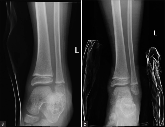
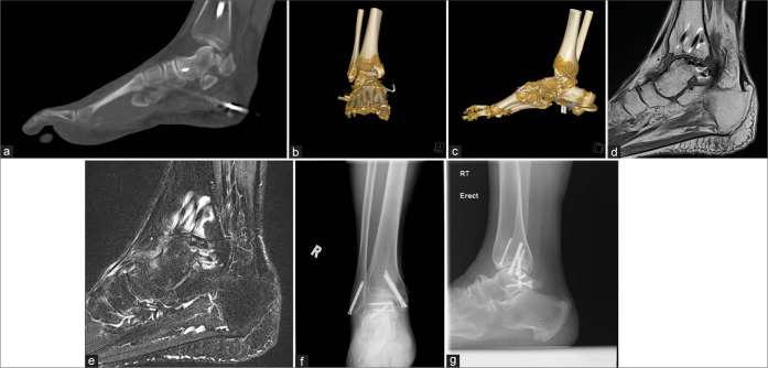
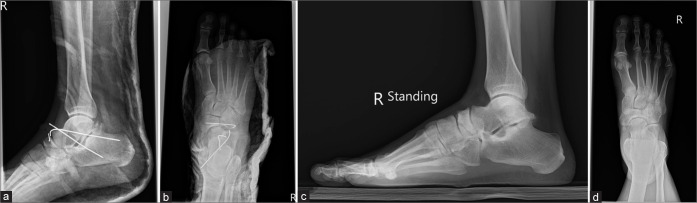
Results: All 23 cases were closed dislocations. Successful close reduction was performed in 17 patients (73.9%) and 6 patients (26.1%) required open reduction and internal fixation. Twenty patients (87%) had associated foot and ankle fractures. Fractures of calcaneal medial tubercle were the most common (75%), followed by talar head (30%), sinus tarsi (25%), and medial malleolus (25%). The radiograph's sensitivity for identifying fractures was 48.1%. The mean follow-up period is 30 months. Symptomatic OA affected 8 patients (36.4%). No post-trauatic talar avascular necrosis was noted. Fractures were present in all of those patients with post-traumatic OA (100%). Three out of five patients who sustained high-energy mechanism injury developed radiographic OA (66.7%). Three out of six patients (50%) treated with open reduction and internal fixation also developed radiographic OA.
Conclusion: Subtalar dislocation remains a rare injury. It is strongly associated with foot and ankle fractures. Fractures of the calcaneal medial tubercle were the most common. The risk of post-traumatic symptomatic peritalar OA is high. CT is useful in detecting occult fractures and injured bony subregions. We postulated potential risk factors of post-traumatic OA (fracture, high-energy mechanism of injury, open reduction, and internal fixation); however, this requires further study.
期刊介绍:
The Journal of Clinical Imaging Science (JCIS) is an open access peer-reviewed journal committed to publishing high-quality articles in the field of Imaging Science. The journal aims to present Imaging Science and relevant clinical information in an understandable and useful format. The journal is owned and published by the Scientific Scholar. Audience Our audience includes Radiologists, Researchers, Clinicians, medical professionals and students. Review process JCIS has a highly rigorous peer-review process that makes sure that manuscripts are scientifically accurate, relevant, novel and important. Authors disclose all conflicts, affiliations and financial associations such that the published content is not biased.

 求助内容:
求助内容: 应助结果提醒方式:


