Sunao Mizumura, Naoyuki Tamamura, Junya Ebina, Hikaru Watanabe, Masaaki Hori
{"title":"Quantitative evaluation of striatal uptake ratios using an adaptive template registration method for 123I-ioflupane dopamine transporter SPECT","authors":"Sunao Mizumura, Naoyuki Tamamura, Junya Ebina, Hikaru Watanabe, Masaaki Hori","doi":"10.1007/s12149-024-01968-8","DOIUrl":null,"url":null,"abstract":"<div><h3>Introduction</h3><p><sup>123</sup>I-FP-CIT (<sup>123</sup>I-Ioflupane) SPECT shows strong accumulation in the striatum, but morphological standardization is challenging due to low accumulation outside the striatum, particularly in subjects with marked striatal decline. In this study, morphological standardization without MRI was achieved using the adaptive template registration (ATR) method to create a subject-specific optimized template with weighted images of normal-type and egg-shape-type templates. The accuracy of a quantitative method for calculating the ratio with nonspecific accumulation in the occipital lobe was evaluated by placing voxels-of-interest (VOI) on standardized images, particularly targeting the striatum.</p><h3>Methods</h3><p>The average images of eight subjects, demonstrating normal-type and egg-shape-type tracer accumulation in <sup>123</sup>I-Ioflupane SPECT, were utilized as normal and disease templates, respectively. The study included 300 subjects that underwent both <sup>123</sup>I-Ioflupane SPECT and MRI for the diagnosis of suspected Parkinson's disease or for exclusion diagnosis. Morphological standardization of SPECT images using structural MRI (MRI-based method) was considered the standard of truth (SOT). Three morphological standardizations without MRI were conducted. The first involved conventional morphological standardization using a normal template (fixed template method), the second employed the ATR method, with a weighted template, and the third used the split-ATR method, processing the left and right striatum separately to address asymmetrical accumulation. VOIs were set on the striatum, caudate, putamen as regions of specific accumulation, and on the occipital lobe as a reference region for nonspecific accumulation.</p><h3>Results</h3><p>Results showed significant and robust linearity in the striatal accumulation ratios for all templates when compared with the occipital lobe accumulation ratio when using the MRI-based method. Comparing intra-class correlations for different linearities, the ATR method and split-ATR method demonstrated higher linearity in the striatum, caudate, and putamen. The split-ATR method showed similar improvements, although more linearity than some of the ATR methods; the effectiveness of the Split-ATR method may vary by image quality, and further validation of its effectiveness in diverse asymmetric accumulation cases seemed warranted.</p><h3>Conclusion</h3><p>The use of optimized templates, such as the ATR and split-ATR methods, improved reproducibility in fully automated processing and demonstrated superior linearity compared to that of MRI-based method, in the ratio to the occipital lobe. The ATR method, which enables morphological standardization when using SPECT images only, proved highly reproducible for clinical quantitative analysis of striatal accumulation, facilitating its clinical use.</p></div>","PeriodicalId":8007,"journal":{"name":"Annals of Nuclear Medicine","volume":"38 12","pages":"943 - 959"},"PeriodicalIF":2.5000,"publicationDate":"2024-08-19","publicationTypes":"Journal Article","fieldsOfStudy":null,"isOpenAccess":false,"openAccessPdf":"https://link.springer.com/content/pdf/10.1007/s12149-024-01968-8.pdf","citationCount":"0","resultStr":null,"platform":"Semanticscholar","paperid":null,"PeriodicalName":"Annals of Nuclear Medicine","FirstCategoryId":"3","ListUrlMain":"https://link.springer.com/article/10.1007/s12149-024-01968-8","RegionNum":4,"RegionCategory":"医学","ArticlePicture":[],"TitleCN":null,"AbstractTextCN":null,"PMCID":null,"EPubDate":"","PubModel":"","JCR":"Q2","JCRName":"RADIOLOGY, NUCLEAR MEDICINE & MEDICAL IMAGING","Score":null,"Total":0}
引用次数: 0
Abstract
Introduction
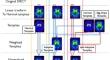
123I-FP-CIT (123I-Ioflupane) SPECT shows strong accumulation in the striatum, but morphological standardization is challenging due to low accumulation outside the striatum, particularly in subjects with marked striatal decline. In this study, morphological standardization without MRI was achieved using the adaptive template registration (ATR) method to create a subject-specific optimized template with weighted images of normal-type and egg-shape-type templates. The accuracy of a quantitative method for calculating the ratio with nonspecific accumulation in the occipital lobe was evaluated by placing voxels-of-interest (VOI) on standardized images, particularly targeting the striatum.
Methods
The average images of eight subjects, demonstrating normal-type and egg-shape-type tracer accumulation in 123I-Ioflupane SPECT, were utilized as normal and disease templates, respectively. The study included 300 subjects that underwent both 123I-Ioflupane SPECT and MRI for the diagnosis of suspected Parkinson's disease or for exclusion diagnosis. Morphological standardization of SPECT images using structural MRI (MRI-based method) was considered the standard of truth (SOT). Three morphological standardizations without MRI were conducted. The first involved conventional morphological standardization using a normal template (fixed template method), the second employed the ATR method, with a weighted template, and the third used the split-ATR method, processing the left and right striatum separately to address asymmetrical accumulation. VOIs were set on the striatum, caudate, putamen as regions of specific accumulation, and on the occipital lobe as a reference region for nonspecific accumulation.
Results
Results showed significant and robust linearity in the striatal accumulation ratios for all templates when compared with the occipital lobe accumulation ratio when using the MRI-based method. Comparing intra-class correlations for different linearities, the ATR method and split-ATR method demonstrated higher linearity in the striatum, caudate, and putamen. The split-ATR method showed similar improvements, although more linearity than some of the ATR methods; the effectiveness of the Split-ATR method may vary by image quality, and further validation of its effectiveness in diverse asymmetric accumulation cases seemed warranted.
Conclusion
The use of optimized templates, such as the ATR and split-ATR methods, improved reproducibility in fully automated processing and demonstrated superior linearity compared to that of MRI-based method, in the ratio to the occipital lobe. The ATR method, which enables morphological standardization when using SPECT images only, proved highly reproducible for clinical quantitative analysis of striatal accumulation, facilitating its clinical use.
期刊介绍:
Annals of Nuclear Medicine is an official journal of the Japanese Society of Nuclear Medicine. It develops the appropriate application of radioactive substances and stable nuclides in the field of medicine.
The journal promotes the exchange of ideas and information and research in nuclear medicine and includes the medical application of radionuclides and related subjects. It presents original articles, short communications, reviews and letters to the editor.

 求助内容:
求助内容: 应助结果提醒方式:


