Feiyu Wei, Xiaohui Kuang, Xi Zhang, Peng Wu, Jie Fan
{"title":"Ventricular activation pattern of left ventricular septal pacing in a canine model.","authors":"Feiyu Wei, Xiaohui Kuang, Xi Zhang, Peng Wu, Jie Fan","doi":"10.1007/s10840-024-01903-x","DOIUrl":null,"url":null,"abstract":"<p><strong>Background: </strong>Left bundle branch pacing (LBBP) is a feasible and effective physiological pacing technique. The QRS morphology of left ventricular septal pacing (LVSP) is similar to that of LBBP. The ventricular activation pattern of LBBP is well-known, whereas the pattern of LVSP still needs further investigation. The present study aimed to determine ventricular activation pattern difference between LVSP and LBBP in a canine model.</p><p><strong>Method: </strong>All six canines underwent successful LBBP and LVSP through trans-ventricular septum using intracardiac echocardiography and intracardiac electrogram. Their hearts were isolated and stained with Lugol's iodine to determine the position of the pacing lead. The activation sequences of the left ventricular myocardium and His-Purkinje system were recorded by placing multiple electrode catheters.</p><p><strong>Results: </strong>First, the left His-Purkinje system in LVSP was activated simultaneously from apical and basal regions to the left ventricular middle septal region, whereas the left ventricular septal myocardium was activated from the apical to basal region. The left His-Purkinje system activation in LBBP occurred in the direction of the apex from the pacing lead, but the left ventricular septal myocardium was activated in the apical to basal direction. Furthermore, the left intraventricular electrical synchrony was similar between LVSP and LBBP as determined by mapping the left ventricular septal to free wall activation time (46.7 ± 1.8 ms vs. 45.0 ± 1.4 ms, p = 0.11).</p><p><strong>Conclusion: </strong>The ventricular activation sequence of LVSP was similar to LBBP. LVSP can capture LBB due to the wide distribution of LBB. These findings suggest a rationale for clinical application of LVSP.</p>","PeriodicalId":16202,"journal":{"name":"Journal of Interventional Cardiac Electrophysiology","volume":" ","pages":"567-577"},"PeriodicalIF":2.6000,"publicationDate":"2025-04-01","publicationTypes":"Journal Article","fieldsOfStudy":null,"isOpenAccess":false,"openAccessPdf":"","citationCount":"0","resultStr":null,"platform":"Semanticscholar","paperid":null,"PeriodicalName":"Journal of Interventional Cardiac Electrophysiology","FirstCategoryId":"3","ListUrlMain":"https://doi.org/10.1007/s10840-024-01903-x","RegionNum":4,"RegionCategory":"医学","ArticlePicture":[],"TitleCN":null,"AbstractTextCN":null,"PMCID":null,"EPubDate":"2024/8/15 0:00:00","PubModel":"Epub","JCR":"Q3","JCRName":"CARDIAC & CARDIOVASCULAR SYSTEMS","Score":null,"Total":0}
引用次数: 0
Abstract
Background: Left bundle branch pacing (LBBP) is a feasible and effective physiological pacing technique. The QRS morphology of left ventricular septal pacing (LVSP) is similar to that of LBBP. The ventricular activation pattern of LBBP is well-known, whereas the pattern of LVSP still needs further investigation. The present study aimed to determine ventricular activation pattern difference between LVSP and LBBP in a canine model.
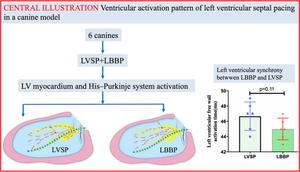
Method: All six canines underwent successful LBBP and LVSP through trans-ventricular septum using intracardiac echocardiography and intracardiac electrogram. Their hearts were isolated and stained with Lugol's iodine to determine the position of the pacing lead. The activation sequences of the left ventricular myocardium and His-Purkinje system were recorded by placing multiple electrode catheters.
Results: First, the left His-Purkinje system in LVSP was activated simultaneously from apical and basal regions to the left ventricular middle septal region, whereas the left ventricular septal myocardium was activated from the apical to basal region. The left His-Purkinje system activation in LBBP occurred in the direction of the apex from the pacing lead, but the left ventricular septal myocardium was activated in the apical to basal direction. Furthermore, the left intraventricular electrical synchrony was similar between LVSP and LBBP as determined by mapping the left ventricular septal to free wall activation time (46.7 ± 1.8 ms vs. 45.0 ± 1.4 ms, p = 0.11).
Conclusion: The ventricular activation sequence of LVSP was similar to LBBP. LVSP can capture LBB due to the wide distribution of LBB. These findings suggest a rationale for clinical application of LVSP.
期刊介绍:
The Journal of Interventional Cardiac Electrophysiology is an international publication devoted to fostering research in and development of interventional techniques and therapies for the management of cardiac arrhythmias. It is designed primarily to present original research studies and scholarly scientific reviews of basic and applied science and clinical research in this field. The Journal will adopt a multidisciplinary approach to link physical, experimental, and clinical sciences as applied to the development of and practice in interventional electrophysiology. The Journal will examine techniques ranging from molecular, chemical and pharmacologic therapies to device and ablation technology. Accordingly, original research in clinical, epidemiologic and basic science arenas will be considered for publication. Applied engineering or physical science studies pertaining to interventional electrophysiology will be encouraged. The Journal is committed to providing comprehensive and detailed treatment of major interventional therapies and innovative techniques in a structured and clinically relevant manner. It is directed at clinical practitioners and investigators in the rapidly growing field of interventional electrophysiology. The editorial staff and board reflect this bias and include noted international experts in this area with a wealth of expertise in basic and clinical investigation. Peer review of all submissions, conflict of interest guidelines and periodic editorial board review of all Journal policies have been established.

 求助内容:
求助内容: 应助结果提醒方式:


