Kacper Stolarz, Aleksander Osiowski, Maciej Preinl, Maksymilian Osiowski, Barbara Jasiewicz, Dominik Taterra
{"title":"The prevalence and anatomy of accessory navicular bone: a meta-analysis.","authors":"Kacper Stolarz, Aleksander Osiowski, Maciej Preinl, Maksymilian Osiowski, Barbara Jasiewicz, Dominik Taterra","doi":"10.1007/s00276-024-03459-x","DOIUrl":null,"url":null,"abstract":"<p><strong>Purpose: </strong>There have been over 40 descriptions of the common developmental variants of the accessory ossicles of the feet. Although predominantly asymptomatic, they sometimes may be linked to painful conditions. One of the most common accessory ossicles in the foot is the accessory navicular bone (AN), located on the medial side of the foot. Our research provides a first meta-analysis on this topic that establishes its frequency by contrasting 39 studies from across the globe.</p><p><strong>Methods: </strong>Up to February 2024, PubMed and Embase databases were thoroughly searched for research on the AN. Eligible data regarding AN prevalence was extracted. This study strictly adhered to the Preferred Reporting Items for Systematic Reviews and Meta-Analyses (PRISMA) guidelines.</p><p><strong>Results: </strong>A total of 39 studies, 11,015 patients, and 36,837 feet were analyzed in our study. The pooled prevalence estimate (PPE) of AN was found to be 17.5% (95%CI: 11.5-25.7) and 12.6% (95%CI: 10.1-15.5) in patients and feet analyses, respectively. Accessory navicular occurred bilaterally in 50.0% of patients, with similar distribution in gender-based groups (21.1% of males and 22.0% of females were confirmed with AN). Accessory navicular was most prevalent in the East Asian population (38.4%) and least prevalent in North Americans (8.0%). No significant differences in AN prevalence were found when comparing different imaging modalities (X-ray and cadaver dissection).</p><p><strong>Conclusion: </strong>Accessory navicular is a common finding in imaging studies. Its prevalence depends on the population covered by the study but is not affected by the patient's gender or the imaging modality utilized for AN assessment.</p>","PeriodicalId":49461,"journal":{"name":"Surgical and Radiologic Anatomy","volume":" ","pages":"1731-1743"},"PeriodicalIF":1.4000,"publicationDate":"2024-10-01","publicationTypes":"Journal Article","fieldsOfStudy":null,"isOpenAccess":false,"openAccessPdf":"https://www.ncbi.nlm.nih.gov/pmc/articles/PMC11405447/pdf/","citationCount":"0","resultStr":null,"platform":"Semanticscholar","paperid":null,"PeriodicalName":"Surgical and Radiologic Anatomy","FirstCategoryId":"3","ListUrlMain":"https://doi.org/10.1007/s00276-024-03459-x","RegionNum":4,"RegionCategory":"医学","ArticlePicture":[],"TitleCN":null,"AbstractTextCN":null,"PMCID":null,"EPubDate":"2024/8/13 0:00:00","PubModel":"Epub","JCR":"Q2","JCRName":"Medicine","Score":null,"Total":0}
引用次数: 0
Abstract
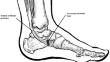
Purpose: There have been over 40 descriptions of the common developmental variants of the accessory ossicles of the feet. Although predominantly asymptomatic, they sometimes may be linked to painful conditions. One of the most common accessory ossicles in the foot is the accessory navicular bone (AN), located on the medial side of the foot. Our research provides a first meta-analysis on this topic that establishes its frequency by contrasting 39 studies from across the globe.
Methods: Up to February 2024, PubMed and Embase databases were thoroughly searched for research on the AN. Eligible data regarding AN prevalence was extracted. This study strictly adhered to the Preferred Reporting Items for Systematic Reviews and Meta-Analyses (PRISMA) guidelines.
Results: A total of 39 studies, 11,015 patients, and 36,837 feet were analyzed in our study. The pooled prevalence estimate (PPE) of AN was found to be 17.5% (95%CI: 11.5-25.7) and 12.6% (95%CI: 10.1-15.5) in patients and feet analyses, respectively. Accessory navicular occurred bilaterally in 50.0% of patients, with similar distribution in gender-based groups (21.1% of males and 22.0% of females were confirmed with AN). Accessory navicular was most prevalent in the East Asian population (38.4%) and least prevalent in North Americans (8.0%). No significant differences in AN prevalence were found when comparing different imaging modalities (X-ray and cadaver dissection).
Conclusion: Accessory navicular is a common finding in imaging studies. Its prevalence depends on the population covered by the study but is not affected by the patient's gender or the imaging modality utilized for AN assessment.
期刊介绍:
Anatomy is a morphological science which cannot fail to interest the clinician. The practical application of anatomical research to clinical problems necessitates special adaptation and selectivity in choosing from numerous international works. Although there is a tendency to believe that meaningful advances in anatomy are unlikely, constant revision is necessary. Surgical and Radiologic Anatomy, the first international journal of Clinical anatomy has been created in this spirit.
Its goal is to serve clinicians, regardless of speciality-physicians, surgeons, radiologists or other specialists-as an indispensable aid with which they can improve their knowledge of anatomy. Each issue includes: Original papers, review articles, articles on the anatomical bases of medical, surgical and radiological techniques, articles of normal radiologic anatomy, brief reviews of anatomical publications of clinical interest.
Particular attention is given to high quality illustrations, which are indispensable for a better understanding of anatomical problems.
Surgical and Radiologic Anatomy is a journal written by anatomists for clinicians with a special interest in anatomy.

 求助内容:
求助内容: 应助结果提醒方式:


