{"title":"Evaluation of primary teeth root canal orifices with naked eye and using magnifying loupes – An in vivo study","authors":"Yamuna Shanmugam , Aksshaya Raghu , M.S. Muthu , Kavitha Swaminathan , Selvakumar Haridoss , K.C. Vignesh , Mayur Bhattad","doi":"10.1016/j.jobcr.2024.08.001","DOIUrl":null,"url":null,"abstract":"<div><h3>Background</h3><p>Knowledge of the anatomy and morphology of root canal orifices and variations are vital elements affecting treatment outcomes.</p></div><div><h3>Aim</h3><p>The objective of this study was to evaluate variations in the number of root canal orifices and their patterns in primary teeth, as identified by both the naked eye and under magnifying loupes.</p></div><div><h3>Materials and methods</h3><p>Total of 173 primary teeth was scheduled for pulpectomy over a period of 18 months. Two examiners assessed the number and pattern of the root canal orifices. After access cavity preparation, the operator recorded the number of root canal orifices with naked eye, and examiner recorded the same using magnifying loupes (3.5×). After cleaning and shaping, the same protocol was used. Collected data were statistically analyzed using SPSS version 23.0 and compared using a paired <em>t</em>-test.</p></div><div><h3>Results</h3><p>The overall variation in the in the identification of root canal orifices between the naked eye and magnifying loupes (3.005 ± 0.971) was statistically significant after access cavity preparation (P ≤ 0.05).</p></div><div><h3>Conclusion</h3><p>Magnifying loupes significantly enhances the determination of the number and pattern of root canal orifices in primary teeth. Therefore, the application of magnifying loupes is essential for accurately assessing variations in root canal orifices in primary dentition.</p></div>","PeriodicalId":16609,"journal":{"name":"Journal of oral biology and craniofacial research","volume":"14 5","pages":"Pages 600-605"},"PeriodicalIF":0.0000,"publicationDate":"2024-08-08","publicationTypes":"Journal Article","fieldsOfStudy":null,"isOpenAccess":false,"openAccessPdf":"https://www.sciencedirect.com/science/article/pii/S2212426824001179/pdfft?md5=e03b0a001d5514856f9f05bac2f42c51&pid=1-s2.0-S2212426824001179-main.pdf","citationCount":"0","resultStr":null,"platform":"Semanticscholar","paperid":null,"PeriodicalName":"Journal of oral biology and craniofacial research","FirstCategoryId":"1085","ListUrlMain":"https://www.sciencedirect.com/science/article/pii/S2212426824001179","RegionNum":0,"RegionCategory":null,"ArticlePicture":[],"TitleCN":null,"AbstractTextCN":null,"PMCID":null,"EPubDate":"","PubModel":"","JCR":"Q1","JCRName":"Medicine","Score":null,"Total":0}
引用次数: 0
Abstract
Background
Knowledge of the anatomy and morphology of root canal orifices and variations are vital elements affecting treatment outcomes.
Aim
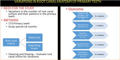
The objective of this study was to evaluate variations in the number of root canal orifices and their patterns in primary teeth, as identified by both the naked eye and under magnifying loupes.
Materials and methods
Total of 173 primary teeth was scheduled for pulpectomy over a period of 18 months. Two examiners assessed the number and pattern of the root canal orifices. After access cavity preparation, the operator recorded the number of root canal orifices with naked eye, and examiner recorded the same using magnifying loupes (3.5×). After cleaning and shaping, the same protocol was used. Collected data were statistically analyzed using SPSS version 23.0 and compared using a paired t-test.
Results
The overall variation in the in the identification of root canal orifices between the naked eye and magnifying loupes (3.005 ± 0.971) was statistically significant after access cavity preparation (P ≤ 0.05).
Conclusion
Magnifying loupes significantly enhances the determination of the number and pattern of root canal orifices in primary teeth. Therefore, the application of magnifying loupes is essential for accurately assessing variations in root canal orifices in primary dentition.
期刊介绍:
Journal of Oral Biology and Craniofacial Research (JOBCR)is the official journal of the Craniofacial Research Foundation (CRF). The journal aims to provide a common platform for both clinical and translational research and to promote interdisciplinary sciences in craniofacial region. JOBCR publishes content that includes diseases, injuries and defects in the head, neck, face, jaws and the hard and soft tissues of the mouth and jaws and face region; diagnosis and medical management of diseases specific to the orofacial tissues and of oral manifestations of systemic diseases; studies on identifying populations at risk of oral disease or in need of specific care, and comparing regional, environmental, social, and access similarities and differences in dental care between populations; diseases of the mouth and related structures like salivary glands, temporomandibular joints, facial muscles and perioral skin; biomedical engineering, tissue engineering and stem cells. The journal publishes reviews, commentaries, peer-reviewed original research articles, short communication, and case reports.

 求助内容:
求助内容: 应助结果提醒方式:


