{"title":"Developing Multichannel smFRET Approach to Dissecting Ribosomal Mechanisms","authors":"Ran Lin, and , Yuhong Wang*, ","doi":"10.1021/cbmi.4c0001010.1021/cbmi.4c00010","DOIUrl":null,"url":null,"abstract":"<p >The ribosome, a 2.6 megadalton biomolecule measuring approximately 20 nm in diameter, coordinates numerous ligands, factors, and regulators to translate proteins with high fidelity and speed. Understanding its complex functions necessitates multiperspective observations. We developed a dual-FRET single-molecule Förste Resonance Energy Transfer method (dual-smFRET), allowing simultaneous observation and correlation of tRNA dynamics and Elongation Factor G (EF-G) conformations in the same complex, in a 10 s time window. By synchronizing laser shutters and motorized filter sets, two FRET signals are captured in consecutive 5 s intervals with a time gap of 50–100 ms. We observed distinct fluorescent emissions from single-, double-, and quadruple-labeled ribosome complexes. Through comprehensive spectrum analysis and correction, we distinguish and correlate conformational changes in two parts of the ribosome, offering additional perspectives on its coordination and timing during translocation. Our setup’s versatility, accommodating up to six FRET pairs, suggests broader applications in studying large biomolecules and various biological systems.</p>","PeriodicalId":53181,"journal":{"name":"Chemical & Biomedical Imaging","volume":"2 7","pages":"501–509 501–509"},"PeriodicalIF":0.0000,"publicationDate":"2024-03-21","publicationTypes":"Journal Article","fieldsOfStudy":null,"isOpenAccess":false,"openAccessPdf":"https://pubs.acs.org/doi/epdf/10.1021/cbmi.4c00010","citationCount":"0","resultStr":null,"platform":"Semanticscholar","paperid":null,"PeriodicalName":"Chemical & Biomedical Imaging","FirstCategoryId":"1085","ListUrlMain":"https://pubs.acs.org/doi/10.1021/cbmi.4c00010","RegionNum":0,"RegionCategory":null,"ArticlePicture":[],"TitleCN":null,"AbstractTextCN":null,"PMCID":null,"EPubDate":"","PubModel":"","JCR":"","JCRName":"","Score":null,"Total":0}
引用次数: 0
Abstract
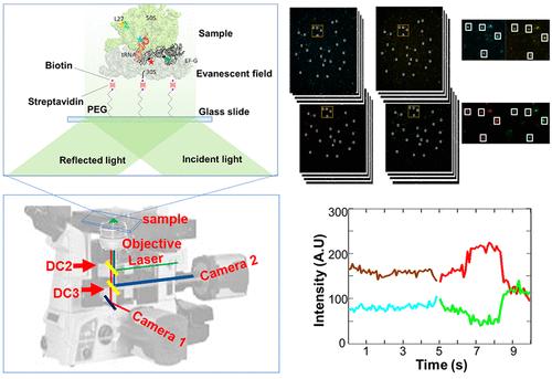
The ribosome, a 2.6 megadalton biomolecule measuring approximately 20 nm in diameter, coordinates numerous ligands, factors, and regulators to translate proteins with high fidelity and speed. Understanding its complex functions necessitates multiperspective observations. We developed a dual-FRET single-molecule Förste Resonance Energy Transfer method (dual-smFRET), allowing simultaneous observation and correlation of tRNA dynamics and Elongation Factor G (EF-G) conformations in the same complex, in a 10 s time window. By synchronizing laser shutters and motorized filter sets, two FRET signals are captured in consecutive 5 s intervals with a time gap of 50–100 ms. We observed distinct fluorescent emissions from single-, double-, and quadruple-labeled ribosome complexes. Through comprehensive spectrum analysis and correction, we distinguish and correlate conformational changes in two parts of the ribosome, offering additional perspectives on its coordination and timing during translocation. Our setup’s versatility, accommodating up to six FRET pairs, suggests broader applications in studying large biomolecules and various biological systems.
期刊介绍:
Chemical & Biomedical Imaging is a peer-reviewed open access journal devoted to the publication of cutting-edge research papers on all aspects of chemical and biomedical imaging. This interdisciplinary field sits at the intersection of chemistry physics biology materials engineering and medicine. The journal aims to bring together researchers from across these disciplines to address cutting-edge challenges of fundamental research and applications.Topics of particular interest include but are not limited to:Imaging of processes and reactionsImaging of nanoscale microscale and mesoscale materialsImaging of biological interactions and interfacesSingle-molecule and cellular imagingWhole-organ and whole-body imagingMolecular imaging probes and contrast agentsBioluminescence chemiluminescence and electrochemiluminescence imagingNanophotonics and imagingChemical tools for new imaging modalitiesChemical and imaging techniques in diagnosis and therapyImaging-guided drug deliveryAI and machine learning assisted imaging
 求助内容:
求助内容: 应助结果提醒方式:


