Apeksha C. Rajamanthrilage, Unaiza Uzair, Paul W. Millhouse, Matthew J. Case, Donald W. Benza and Jeffrey N. Anker*,
{"title":"Spatial Resolution for X-ray Excited Luminescence Chemical Imaging (XELCI)","authors":"Apeksha C. Rajamanthrilage, Unaiza Uzair, Paul W. Millhouse, Matthew J. Case, Donald W. Benza and Jeffrey N. Anker*, ","doi":"10.1021/cbmi.4c0003910.1021/cbmi.4c00039","DOIUrl":null,"url":null,"abstract":"<p >Measuring chemical concentrations at the surface of implanted medical devices is important for elucidating the local biochemical environment, especially during implant infection. Although chemical indicator dyes enable chemical measurements in vitro, they are usually ineffective when measuring through tissue because the background obscures the dye signal and scattering dramatically reduces the spatial resolution. X-ray excited luminescent chemical imaging (XELCI) is a recent imaging modality which overcomes these limitations using a focused X-ray beam to excite a small spot of red light on scintillator-coated medical implants with well-defined location (because X-rays are minimally scattered) and low background. A spectrochemical indicator film placed over the scintillator layer, e.g., a polymer film containing pH-indicator dyes, absorbs some of the luminescence according to the local chemical environment, and this absorption is then detected by measuring the light intensity/spectrum passing through the tissue. A focused X-ray beam is used to scan point-by-point with a spatial resolution mainly limited by the X-ray beam width with minimum increase from X-ray absorption and scattering in the tissue. X-ray resolution, implant surface specificity, and chemical sensitivity are the three key features of XELCI. Here, we study spatial resolution using optically absorptive targets. For imaging a series of lines, the 20–80% knife-edge resolution was ∼285 (±15) μm with no tissue and 475 ± 18 and 520 ± 34 μm, respectively, through 5 and 10 mm thick tissue. Thus, doubling the tissue depth did not appreciably change the spatial resolution recorded through the tissue. This shows the promise of XELCI for submillimeter chemical imaging through tissue.</p>","PeriodicalId":53181,"journal":{"name":"Chemical & Biomedical Imaging","volume":"2 7","pages":"510–517 510–517"},"PeriodicalIF":0.0000,"publicationDate":"2024-07-02","publicationTypes":"Journal Article","fieldsOfStudy":null,"isOpenAccess":false,"openAccessPdf":"https://pubs.acs.org/doi/epdf/10.1021/cbmi.4c00039","citationCount":"0","resultStr":null,"platform":"Semanticscholar","paperid":null,"PeriodicalName":"Chemical & Biomedical Imaging","FirstCategoryId":"1085","ListUrlMain":"https://pubs.acs.org/doi/10.1021/cbmi.4c00039","RegionNum":0,"RegionCategory":null,"ArticlePicture":[],"TitleCN":null,"AbstractTextCN":null,"PMCID":null,"EPubDate":"","PubModel":"","JCR":"","JCRName":"","Score":null,"Total":0}
引用次数: 0
Abstract
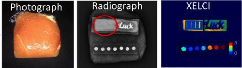
Measuring chemical concentrations at the surface of implanted medical devices is important for elucidating the local biochemical environment, especially during implant infection. Although chemical indicator dyes enable chemical measurements in vitro, they are usually ineffective when measuring through tissue because the background obscures the dye signal and scattering dramatically reduces the spatial resolution. X-ray excited luminescent chemical imaging (XELCI) is a recent imaging modality which overcomes these limitations using a focused X-ray beam to excite a small spot of red light on scintillator-coated medical implants with well-defined location (because X-rays are minimally scattered) and low background. A spectrochemical indicator film placed over the scintillator layer, e.g., a polymer film containing pH-indicator dyes, absorbs some of the luminescence according to the local chemical environment, and this absorption is then detected by measuring the light intensity/spectrum passing through the tissue. A focused X-ray beam is used to scan point-by-point with a spatial resolution mainly limited by the X-ray beam width with minimum increase from X-ray absorption and scattering in the tissue. X-ray resolution, implant surface specificity, and chemical sensitivity are the three key features of XELCI. Here, we study spatial resolution using optically absorptive targets. For imaging a series of lines, the 20–80% knife-edge resolution was ∼285 (±15) μm with no tissue and 475 ± 18 and 520 ± 34 μm, respectively, through 5 and 10 mm thick tissue. Thus, doubling the tissue depth did not appreciably change the spatial resolution recorded through the tissue. This shows the promise of XELCI for submillimeter chemical imaging through tissue.
期刊介绍:
Chemical & Biomedical Imaging is a peer-reviewed open access journal devoted to the publication of cutting-edge research papers on all aspects of chemical and biomedical imaging. This interdisciplinary field sits at the intersection of chemistry physics biology materials engineering and medicine. The journal aims to bring together researchers from across these disciplines to address cutting-edge challenges of fundamental research and applications.Topics of particular interest include but are not limited to:Imaging of processes and reactionsImaging of nanoscale microscale and mesoscale materialsImaging of biological interactions and interfacesSingle-molecule and cellular imagingWhole-organ and whole-body imagingMolecular imaging probes and contrast agentsBioluminescence chemiluminescence and electrochemiluminescence imagingNanophotonics and imagingChemical tools for new imaging modalitiesChemical and imaging techniques in diagnosis and therapyImaging-guided drug deliveryAI and machine learning assisted imaging
 求助内容:
求助内容: 应助结果提醒方式:


