{"title":"Continuum topological derivative - A novel application tool for segmentation of CT and MRI images","authors":"Viswanath Muthukrishnan , Sandeep Jaipurkar , Nedumaran Damodaran","doi":"10.1016/j.ynirp.2024.100215","DOIUrl":null,"url":null,"abstract":"<div><h3>Introduction</h3><p>Computed Tomography (CT) and Magnetic Resonance Imaging (MRI) are essential tools for unraveling anatomical and tissue properties, particularly in the head and brain. CT provides high-contrast images, particularly valuable in cases such as cerebral bleeds, and also aids in estimating cranial deformities and organ shape deviations. MRI, on the other hand, offers excellent imaging of cerebral artery regions, allowing analysis of various cerebral pathologies through different sequences. Beyond detecting common head and brain disorders, these modalities play a crucial role in identifying abnormalities in orbits, middle cerebral artery territories, brain ventricles, soft tissues, and bones. A unique aspect of brain MRI is its ability to produce multiplanar brain assessments. Both head/brain CT and MRI are invaluable for studying haemorrhage cases, with segmentation of affected areas providing detailed images for further analysis. This study explores the application of a novel mathematical technique, continuum topological derivative (CTD), for CT and MR image segmentation.</p></div><div><h3>Methods</h3><p>The initial stage of Continuum Topological Derivative (CTD) segmentation involves preprocessing CT and MR images due to their susceptibility to inherent noises, such as quantum mottle, and Gaussian and Rayleigh noises, respectively. In this study, we have implemented the CTD denoising algorithm to produce denoised CT/MR images, serving as ground truth for subsequent segmentation steps. Validation of the denoised CTD CT/MR images was conducted through minimal residual value computation across all case studies. Following this, segmentation of the region of interest was performed using the CTD technique, with comparisons made against Discrete Topological Derivatives (DTD), k-mean clustering and Adaptive Threshold methods. Evaluation of the proposed CTD algorithm's effectiveness in segmentation involved calculating performance metrics such as Jaccard and dice indices to assess spatial overlap of segmented images.</p></div><div><h3>Results</h3><p>The CTD technique yields excellent segmentation results, not only for the delineated region of interest but also for volume-based cerebral blood areas and anomalies in the middle cerebral artery (MCA) and its territorial areas, which are substantiated through performance metrics and visual inspection by trained radiologist. This aids in determining the severity of stroke in affected patients. Additionally, a unique attempt is made to apply CTD to Electrical Impedance Tomography (EIT) images of the lungs for precise estimation of the breathing cycle. CTD successfully generates standardized images, demonstrating attenuation and density characteristics for cerebral cisterns, arteries, and ventricles.</p></div><div><h3>Discussion</h3><p>The denoised images obtained through CTD facilitate thorough analysis of both normal and pathological conditions, providing radiologists with enhanced capabilities to identify subtle details, particularly in areas such as abnormal cerebral artery territories, haemorrhage cases, cisterns, ventricles and arteries. Results clearly demonstrate that the combination of CTD denoising and segmentation outperforms the other three established methods in terms of both efficiency and accuracy in delineating diseased or affected areas, as evidenced by the various case studies conducted in this research. In summary, the proposed CTD method aims to delineate boundaries and contours of the region of interest, facilitating precise estimation of size and shape for accurate detection of the extent of diseased or affected areas.</p></div>","PeriodicalId":74277,"journal":{"name":"Neuroimage. Reports","volume":"4 3","pages":"Article 100215"},"PeriodicalIF":0.0000,"publicationDate":"2024-08-01","publicationTypes":"Journal Article","fieldsOfStudy":null,"isOpenAccess":false,"openAccessPdf":"https://www.sciencedirect.com/science/article/pii/S2666956024000217/pdfft?md5=e00786ad1e1b5c9b31d74350ad01cd38&pid=1-s2.0-S2666956024000217-main.pdf","citationCount":"0","resultStr":null,"platform":"Semanticscholar","paperid":null,"PeriodicalName":"Neuroimage. Reports","FirstCategoryId":"1085","ListUrlMain":"https://www.sciencedirect.com/science/article/pii/S2666956024000217","RegionNum":0,"RegionCategory":null,"ArticlePicture":[],"TitleCN":null,"AbstractTextCN":null,"PMCID":null,"EPubDate":"","PubModel":"","JCR":"Q4","JCRName":"Neuroscience","Score":null,"Total":0}
引用次数: 0
Abstract
Introduction
Computed Tomography (CT) and Magnetic Resonance Imaging (MRI) are essential tools for unraveling anatomical and tissue properties, particularly in the head and brain. CT provides high-contrast images, particularly valuable in cases such as cerebral bleeds, and also aids in estimating cranial deformities and organ shape deviations. MRI, on the other hand, offers excellent imaging of cerebral artery regions, allowing analysis of various cerebral pathologies through different sequences. Beyond detecting common head and brain disorders, these modalities play a crucial role in identifying abnormalities in orbits, middle cerebral artery territories, brain ventricles, soft tissues, and bones. A unique aspect of brain MRI is its ability to produce multiplanar brain assessments. Both head/brain CT and MRI are invaluable for studying haemorrhage cases, with segmentation of affected areas providing detailed images for further analysis. This study explores the application of a novel mathematical technique, continuum topological derivative (CTD), for CT and MR image segmentation.
Methods
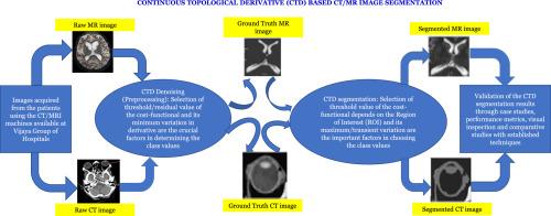
The initial stage of Continuum Topological Derivative (CTD) segmentation involves preprocessing CT and MR images due to their susceptibility to inherent noises, such as quantum mottle, and Gaussian and Rayleigh noises, respectively. In this study, we have implemented the CTD denoising algorithm to produce denoised CT/MR images, serving as ground truth for subsequent segmentation steps. Validation of the denoised CTD CT/MR images was conducted through minimal residual value computation across all case studies. Following this, segmentation of the region of interest was performed using the CTD technique, with comparisons made against Discrete Topological Derivatives (DTD), k-mean clustering and Adaptive Threshold methods. Evaluation of the proposed CTD algorithm's effectiveness in segmentation involved calculating performance metrics such as Jaccard and dice indices to assess spatial overlap of segmented images.
Results
The CTD technique yields excellent segmentation results, not only for the delineated region of interest but also for volume-based cerebral blood areas and anomalies in the middle cerebral artery (MCA) and its territorial areas, which are substantiated through performance metrics and visual inspection by trained radiologist. This aids in determining the severity of stroke in affected patients. Additionally, a unique attempt is made to apply CTD to Electrical Impedance Tomography (EIT) images of the lungs for precise estimation of the breathing cycle. CTD successfully generates standardized images, demonstrating attenuation and density characteristics for cerebral cisterns, arteries, and ventricles.
Discussion
The denoised images obtained through CTD facilitate thorough analysis of both normal and pathological conditions, providing radiologists with enhanced capabilities to identify subtle details, particularly in areas such as abnormal cerebral artery territories, haemorrhage cases, cisterns, ventricles and arteries. Results clearly demonstrate that the combination of CTD denoising and segmentation outperforms the other three established methods in terms of both efficiency and accuracy in delineating diseased or affected areas, as evidenced by the various case studies conducted in this research. In summary, the proposed CTD method aims to delineate boundaries and contours of the region of interest, facilitating precise estimation of size and shape for accurate detection of the extent of diseased or affected areas.

 求助内容:
求助内容: 应助结果提醒方式:


