Maria Correia de Verdier, Elisabeth Ronne-Engström, Ljubisa Borota, Johan Wikström
{"title":"Hemodynamic evaluation of intracranial arteriovenous malformations: Pre- and post-treatment 2D phase-contrast MRI measurements.","authors":"Maria Correia de Verdier, Elisabeth Ronne-Engström, Ljubisa Borota, Johan Wikström","doi":"10.1177/20584601241269608","DOIUrl":null,"url":null,"abstract":"<p><strong>Background: </strong>Hemodynamic changes are seen in the feeding arteries of arteriovenous malformations (AVMs). Phase-contrast MRI (PC-MRI) enables the acquisition of hemodynamic information from blood vessels. There is insufficient knowledge on which flow or velocity parameter best discriminates AVMs from healthy subjects.</p><p><strong>Purpose: </strong>To evaluate PC-MRI-measured flow and velocity in feeding arteries of AVMs before and, when possible, also after treatment and to compare these measurements to corresponding measurements in healthy controls.</p><p><strong>Materials and methods: </strong>Highest flow (HF), lowest flow (LF), mean flow (MF), peak systolic velocity (PSV), end-diastolic velocity (EDV), and mean velocity (MV) were measured in feeding arteries in patients with intracranial AVMs using 2D PC-MRI at 3 T. Measurements were compared to previously reported values in healthy individuals. Values in patients above the 95th percentile in the healthy cohort were categorized as pathological. Nidus volume was measured using 3D time-of-flight MR angiography.</p><p><strong>Results: </strong>Ten patients with diagnosed AVMs were examined with PC-MRI. Among these, three patients also underwent follow-up PC-MRI after treatment. Pathological velocities (PSV, EDV, and MV) were seen in all five subjects with a nidus larger or equal to 5.7 cm<sup>3</sup>, whereas pathological flow values were not seen in all, that is, pathologic HF in three, pathologic LF in two, and pathologic MF in two. After treatment, there was a decrease in flow and velocity (all measured parameters). After treatment, velocities (PSV, EDV, and MV) were no longer abnormal compared to healthy controls.</p><p><strong>Conclusion: </strong>Patients with a large AVM nidus show pathological velocities, but less consistent flow increases. Following treatment, velocities normalize.</p>","PeriodicalId":72063,"journal":{"name":"Acta radiologica open","volume":"13 8","pages":"20584601241269608"},"PeriodicalIF":1.0000,"publicationDate":"2024-08-08","publicationTypes":"Journal Article","fieldsOfStudy":null,"isOpenAccess":false,"openAccessPdf":"https://www.ncbi.nlm.nih.gov/pmc/articles/PMC11311173/pdf/","citationCount":"0","resultStr":null,"platform":"Semanticscholar","paperid":null,"PeriodicalName":"Acta radiologica open","FirstCategoryId":"1085","ListUrlMain":"https://doi.org/10.1177/20584601241269608","RegionNum":0,"RegionCategory":null,"ArticlePicture":[],"TitleCN":null,"AbstractTextCN":null,"PMCID":null,"EPubDate":"2024/8/1 0:00:00","PubModel":"eCollection","JCR":"Q4","JCRName":"RADIOLOGY, NUCLEAR MEDICINE & MEDICAL IMAGING","Score":null,"Total":0}
引用次数: 0
Abstract
Background: Hemodynamic changes are seen in the feeding arteries of arteriovenous malformations (AVMs). Phase-contrast MRI (PC-MRI) enables the acquisition of hemodynamic information from blood vessels. There is insufficient knowledge on which flow or velocity parameter best discriminates AVMs from healthy subjects.
Purpose: To evaluate PC-MRI-measured flow and velocity in feeding arteries of AVMs before and, when possible, also after treatment and to compare these measurements to corresponding measurements in healthy controls.
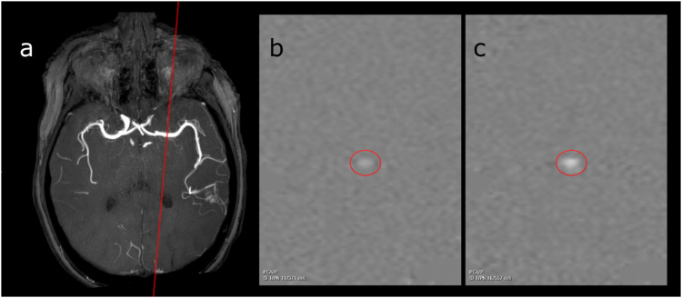
Materials and methods: Highest flow (HF), lowest flow (LF), mean flow (MF), peak systolic velocity (PSV), end-diastolic velocity (EDV), and mean velocity (MV) were measured in feeding arteries in patients with intracranial AVMs using 2D PC-MRI at 3 T. Measurements were compared to previously reported values in healthy individuals. Values in patients above the 95th percentile in the healthy cohort were categorized as pathological. Nidus volume was measured using 3D time-of-flight MR angiography.
Results: Ten patients with diagnosed AVMs were examined with PC-MRI. Among these, three patients also underwent follow-up PC-MRI after treatment. Pathological velocities (PSV, EDV, and MV) were seen in all five subjects with a nidus larger or equal to 5.7 cm3, whereas pathological flow values were not seen in all, that is, pathologic HF in three, pathologic LF in two, and pathologic MF in two. After treatment, there was a decrease in flow and velocity (all measured parameters). After treatment, velocities (PSV, EDV, and MV) were no longer abnormal compared to healthy controls.
Conclusion: Patients with a large AVM nidus show pathological velocities, but less consistent flow increases. Following treatment, velocities normalize.

 求助内容:
求助内容: 应助结果提醒方式:


