{"title":"Feasibility of a single-phase portal venous CT protocol using bolus tracking technique and lean body weight-based contrast media dose.","authors":"Riccardo Valletta, Matteo Bonatti, Vincenzo Vingiani, Valentina Corato, Bernardo Proner, Fabio Lombardo, Giacomo Avesani, Patrizia Pertner, Giulia A Zamboni","doi":"10.1007/s00330-024-11009-7","DOIUrl":null,"url":null,"abstract":"<p><strong>Purpose: </strong>To evaluate the impact of the use of lean body weight (LBW)-based contrast material (CM) dose and bolus tracking technique on portal venous phase abdominal CT image quality.</p><p><strong>Materials and methods: </strong>IRB-approved prospective study; informed consent was acquired. In the period July-November 2023, we randomly selected 105 oncologic patients scheduled for a portal venous phase abdominal CT to undergo our experimental protocol (i.e., 0.7 gI/Kg of LBW CM administration and bolus tracking on the liver). Included patients had performed a \"standard\" portal venous phase abdominal CT (i.e., 0.6 gI/Kg of total body weight (TBW) contrast material administration and 70 s fixed delay) on the same scanner within the previous 12 months. One reader evaluated CT images measuring liver, portal vein, kidney cortex, and spleen attenuation; values were normalized to paraspinal muscles.</p><p><strong>Results: </strong>Median administered contrast dose (350 mgI/mL CM) was 99 mL (IQR: 81-115 mL) using the experimental protocol and 110 mL (IQR: 100-120 mL) using the standard one (p < 0.0001). Median acquisition delay using the experimental protocol was 65\" (IQR 59-73\"). Median normalized hepatic enhancement was significantly higher using the experimental protocol (1.97, IQR: 1.83-2.47 vs. 1.86, IQR: 1.58-2.11; p < 0.0001). Median normalized portal vein enhancement was significantly higher using the experimental protocol (3.43, IQR: 2.73-4.04 vs. 2.91, IQR: 2.58-3.41; p < 0.0001). No statistically significant differences were found in the kidneys' cortex and aorta normalized enhancement (p > 0.05).</p><p><strong>Conclusion: </strong>The combination of LBW-based CM dose administration and bolus tracking allows a significant CM dose reduction and a significant liver and portal vein enhancement increase.</p><p><strong>Clinical relevance statement: </strong>Lean body weight-based contrast material (CM) dose administration and bolus tracking technique in portal venous phase CT scans overcome differences in body composition and hemodynamics, improving reproducibility. It allows a significant CM dose reduction with increased liver and portal vein enhancement.</p><p><strong>Key points: </strong>Lean body weight (LBW)-based contrast material (CM) dosing could be superior to total body weight dosing. Portal venous phase CT with a liver bolus tracking technique improved liver and spleen enhancement with a reduced contrast dose. The combination of LBW-based CM dosing and liver bolus tracking technique enables more \"customized\" CT examinations.</p>","PeriodicalId":12076,"journal":{"name":"European Radiology","volume":" ","pages":"1067-1075"},"PeriodicalIF":4.7000,"publicationDate":"2025-02-01","publicationTypes":"Journal Article","fieldsOfStudy":null,"isOpenAccess":false,"openAccessPdf":"","citationCount":"0","resultStr":null,"platform":"Semanticscholar","paperid":null,"PeriodicalName":"European Radiology","FirstCategoryId":"3","ListUrlMain":"https://doi.org/10.1007/s00330-024-11009-7","RegionNum":2,"RegionCategory":"医学","ArticlePicture":[],"TitleCN":null,"AbstractTextCN":null,"PMCID":null,"EPubDate":"2024/8/9 0:00:00","PubModel":"Epub","JCR":"Q1","JCRName":"RADIOLOGY, NUCLEAR MEDICINE & MEDICAL IMAGING","Score":null,"Total":0}
引用次数: 0
Abstract
Purpose: To evaluate the impact of the use of lean body weight (LBW)-based contrast material (CM) dose and bolus tracking technique on portal venous phase abdominal CT image quality.
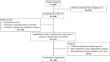
Materials and methods: IRB-approved prospective study; informed consent was acquired. In the period July-November 2023, we randomly selected 105 oncologic patients scheduled for a portal venous phase abdominal CT to undergo our experimental protocol (i.e., 0.7 gI/Kg of LBW CM administration and bolus tracking on the liver). Included patients had performed a "standard" portal venous phase abdominal CT (i.e., 0.6 gI/Kg of total body weight (TBW) contrast material administration and 70 s fixed delay) on the same scanner within the previous 12 months. One reader evaluated CT images measuring liver, portal vein, kidney cortex, and spleen attenuation; values were normalized to paraspinal muscles.
Results: Median administered contrast dose (350 mgI/mL CM) was 99 mL (IQR: 81-115 mL) using the experimental protocol and 110 mL (IQR: 100-120 mL) using the standard one (p < 0.0001). Median acquisition delay using the experimental protocol was 65" (IQR 59-73"). Median normalized hepatic enhancement was significantly higher using the experimental protocol (1.97, IQR: 1.83-2.47 vs. 1.86, IQR: 1.58-2.11; p < 0.0001). Median normalized portal vein enhancement was significantly higher using the experimental protocol (3.43, IQR: 2.73-4.04 vs. 2.91, IQR: 2.58-3.41; p < 0.0001). No statistically significant differences were found in the kidneys' cortex and aorta normalized enhancement (p > 0.05).
Conclusion: The combination of LBW-based CM dose administration and bolus tracking allows a significant CM dose reduction and a significant liver and portal vein enhancement increase.
Clinical relevance statement: Lean body weight-based contrast material (CM) dose administration and bolus tracking technique in portal venous phase CT scans overcome differences in body composition and hemodynamics, improving reproducibility. It allows a significant CM dose reduction with increased liver and portal vein enhancement.
Key points: Lean body weight (LBW)-based contrast material (CM) dosing could be superior to total body weight dosing. Portal venous phase CT with a liver bolus tracking technique improved liver and spleen enhancement with a reduced contrast dose. The combination of LBW-based CM dosing and liver bolus tracking technique enables more "customized" CT examinations.
期刊介绍:
European Radiology (ER) continuously updates scientific knowledge in radiology by publication of strong original articles and state-of-the-art reviews written by leading radiologists. A well balanced combination of review articles, original papers, short communications from European radiological congresses and information on society matters makes ER an indispensable source for current information in this field.
This is the Journal of the European Society of Radiology, and the official journal of a number of societies.
From 2004-2008 supplements to European Radiology were published under its companion, European Radiology Supplements, ISSN 1613-3749.

 求助内容:
求助内容: 应助结果提醒方式:


