{"title":"Choroidal vascular changes in early-stage myopic maculopathy from deep learning choroidal analysis: a hospital-based SS-OCT study.","authors":"Yan Li, Haoer Li, Xue Rui, Yuan Wang, Shenju Zhu, Mengge Huang, Jianqiang Liang, Yangfeifei Zhu, Jiajia Shi, Le Yu, Shenghai Huang, Chun Yang, Mengmeng Dong, Hebei Gao, Meixiao Shen, Hao Wu, Xiangtian Zhou","doi":"10.1186/s40662-024-00398-x","DOIUrl":null,"url":null,"abstract":"<p><strong>Background: </strong>The objective of this study is to illustrate the changes in the choroidal vasculature in individuals with diffuse chorioretinal atrophy (DCA, early-stage myopic maculopathy) and investigate the association between them.</p><p><strong>Methods: </strong>This study included 1418 highly myopic eyes from 720 participants aged 18 - 60 years from the Wenzhou High Myopia Cohort Study. These participants underwent comprehensive ophthalmic assessments. Myopic maculopathy classification followed the Meta-PM system, with pathological myopia defined as myopic maculopathy of DCA or severer. Eyes with myopic maculopathy categorized as no macular lesions (C0), tessellated fundus (C1), and DCA (C2) were enrolled in the analysis. Choroidal images were obtained from swept-source optical coherence tomography (SS-OCT), and the images were processed with a deep learning-based automatic segmentation algorithm and the Niblack auto-local threshold algorithm.</p><p><strong>Results: </strong>DCA was detected in 247 eyes (17.4%). In comparison to eyes with C0, those with C2 exhibited significant reductions in choroidal thickness (ChT), luminal area (LA), and stromal area (SA) across all evaluated regions (all P < 0.001). An increase in choroidal vascular index (CVI) was observed in all regions, except for the nasal perifoveal (N2) and inferior perifoveal (I2) regions (all P < 0.01). Multivariable logistic regression analysis revealed a negative association between the presence of DCA and increases in choroidal LA and SA (odds ratio ≤ 0.099, P < 0.001). Multivariable linear regression analysis showed that the mean deviation of the visual field test was positively associated with LA and SA at the vertical meridian (B = 1.512, P < 0.001 for LA; B = 1.956, P < 0.001 for SA). Furthermore, the receiver operating characteristic curve analyses showed the optimal ChT to diagnose pathological myopia was 82.4 µm in the N2 region, the LA was 0.076 mm<sup>2</sup> and the SA was 0.049 mm<sup>2</sup>, with area under the curves of 0.916, 0.908, and 0.895, respectively.</p><p><strong>Conclusions: </strong>The results of this study indicated that both the presence of DCA and visual function impairment were associated with reductions in choroidal perfusion and stromal components. Moreover, we established threshold values for choroidal parameters in diagnosing pathological myopia, offering valuable references for clinical diagnosis and management.</p>","PeriodicalId":12194,"journal":{"name":"Eye and Vision","volume":"11 1","pages":"32"},"PeriodicalIF":4.0000,"publicationDate":"2024-08-06","publicationTypes":"Journal Article","fieldsOfStudy":null,"isOpenAccess":false,"openAccessPdf":"https://www.ncbi.nlm.nih.gov/pmc/articles/PMC11301841/pdf/","citationCount":"0","resultStr":null,"platform":"Semanticscholar","paperid":null,"PeriodicalName":"Eye and Vision","FirstCategoryId":"3","ListUrlMain":"https://doi.org/10.1186/s40662-024-00398-x","RegionNum":1,"RegionCategory":"医学","ArticlePicture":[],"TitleCN":null,"AbstractTextCN":null,"PMCID":null,"EPubDate":"","PubModel":"","JCR":"Q1","JCRName":"OPHTHALMOLOGY","Score":null,"Total":0}
引用次数: 0
Abstract
Background: The objective of this study is to illustrate the changes in the choroidal vasculature in individuals with diffuse chorioretinal atrophy (DCA, early-stage myopic maculopathy) and investigate the association between them.
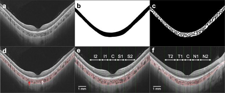
Methods: This study included 1418 highly myopic eyes from 720 participants aged 18 - 60 years from the Wenzhou High Myopia Cohort Study. These participants underwent comprehensive ophthalmic assessments. Myopic maculopathy classification followed the Meta-PM system, with pathological myopia defined as myopic maculopathy of DCA or severer. Eyes with myopic maculopathy categorized as no macular lesions (C0), tessellated fundus (C1), and DCA (C2) were enrolled in the analysis. Choroidal images were obtained from swept-source optical coherence tomography (SS-OCT), and the images were processed with a deep learning-based automatic segmentation algorithm and the Niblack auto-local threshold algorithm.
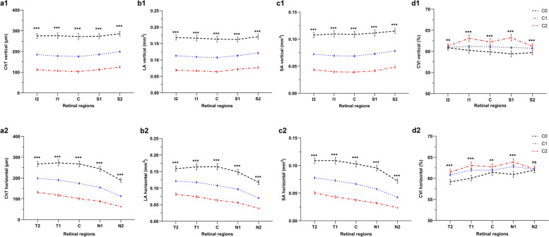
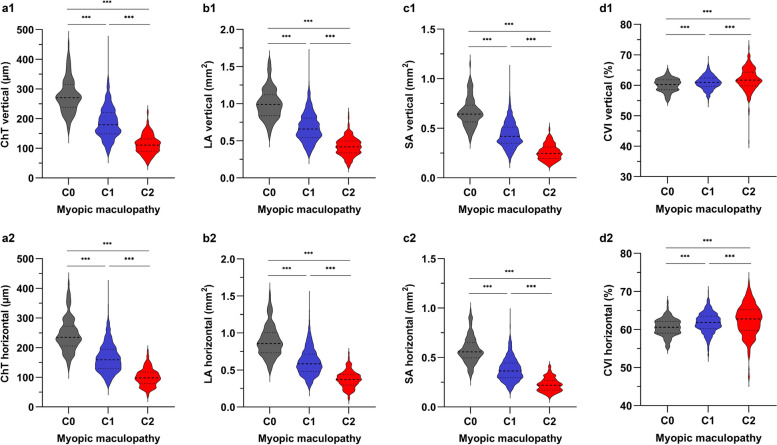
Results: DCA was detected in 247 eyes (17.4%). In comparison to eyes with C0, those with C2 exhibited significant reductions in choroidal thickness (ChT), luminal area (LA), and stromal area (SA) across all evaluated regions (all P < 0.001). An increase in choroidal vascular index (CVI) was observed in all regions, except for the nasal perifoveal (N2) and inferior perifoveal (I2) regions (all P < 0.01). Multivariable logistic regression analysis revealed a negative association between the presence of DCA and increases in choroidal LA and SA (odds ratio ≤ 0.099, P < 0.001). Multivariable linear regression analysis showed that the mean deviation of the visual field test was positively associated with LA and SA at the vertical meridian (B = 1.512, P < 0.001 for LA; B = 1.956, P < 0.001 for SA). Furthermore, the receiver operating characteristic curve analyses showed the optimal ChT to diagnose pathological myopia was 82.4 µm in the N2 region, the LA was 0.076 mm2 and the SA was 0.049 mm2, with area under the curves of 0.916, 0.908, and 0.895, respectively.
Conclusions: The results of this study indicated that both the presence of DCA and visual function impairment were associated with reductions in choroidal perfusion and stromal components. Moreover, we established threshold values for choroidal parameters in diagnosing pathological myopia, offering valuable references for clinical diagnosis and management.
期刊介绍:
Eye and Vision is an open access, peer-reviewed journal for ophthalmologists and visual science specialists. It welcomes research articles, reviews, methodologies, commentaries, case reports, perspectives and short reports encompassing all aspects of eye and vision. Topics of interest include but are not limited to: current developments of theoretical, experimental and clinical investigations in ophthalmology, optometry and vision science which focus on novel and high-impact findings on central issues pertaining to biology, pathophysiology and etiology of eye diseases as well as advances in diagnostic techniques, surgical treatment, instrument updates, the latest drug findings, results of clinical trials and research findings. It aims to provide ophthalmologists and visual science specialists with the latest developments in theoretical, experimental and clinical investigations in eye and vision.
 求助内容:
求助内容: 应助结果提醒方式:


